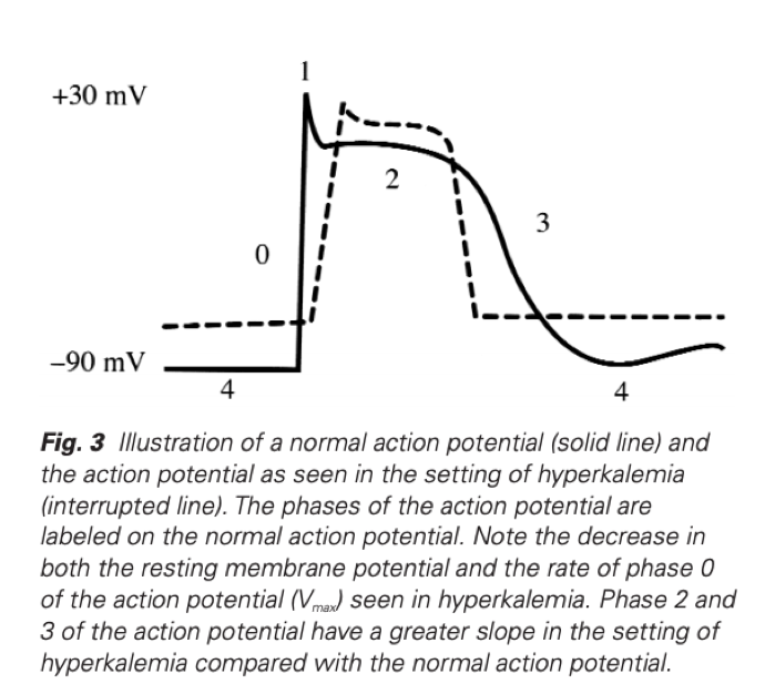
Velocity (Vmax) of Ph 0 decreases as RMP becomes less negative
Threshold potential decreases (from -75mV to -70mV)
Increase in slope of Ph 2 & 3 shortening repolarisation time (responsible for ST-T depression, peaked Tw & QT shortening)
Hyperkalaemia Revisited, Parham W et al, Tex Heart Inst J 2006;33:40-7
3.
The position with the greatest reduction in FRC:
Supine
Trendelenburg
Erect
Prone
4.
The correct order of the ECG and CVP trace on Wigger’s diagram:
C wave R wave T wave V wave
R wave C wave V wave T wave
C wave R wave T wave V wave
R wave C wave T wave V wave
5.
Capacitance is measured in
Volts
Watts
Farads
Joules
This qn was recalled as something in regards to a defibrillator capacitance refers to…Capacitance is the ability to store charge (SI = Farad)
6.
The correct surface anatomy of the radial artery in the distal forearm
Medial to the Median nerve
Lateral to the flexor carpi radialis
Medial to the Ulnar nerve
Lateral to the brachioradialis
7.
The most lateral structure:
Femoral Artery
Femoral Vein
Femoral Nerve
Femoral Canal
Too easy.They could potentially confuse by asking you the most lateral structure in the femoral sheath (as opposed to the triangle which does not contain the nerve)
8.
The events occurring in Phase II of the Valsalva Manoeuvre:
↓BP, ↑HR, ↑Intrathoracic Pressure
↑BP, ↓HR, ↑Intrathoracic Pressure
↓BP, ↑HR, ↓Intrathoracic Pressure
↑BP, ↓HR, ↓Intrathoracic Pressure
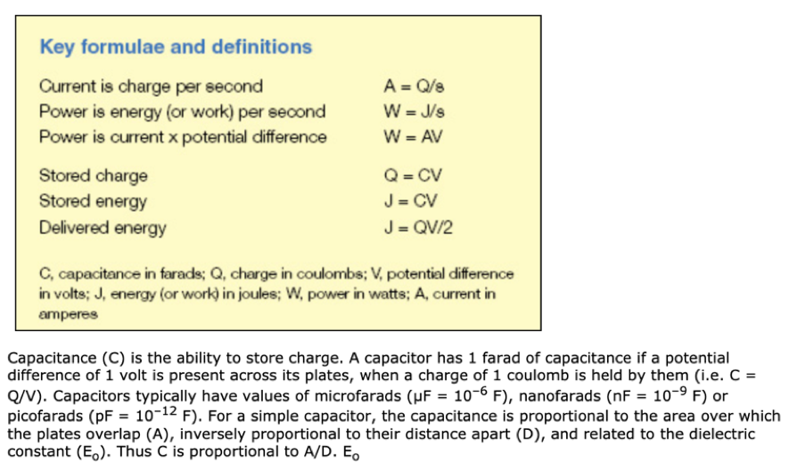
An additional variable added to the 2019 MCQ
9.
Permanent closure of Ductus Arteriosus after birth occurs
1-5 mins
1-5 hours
2-3 days
2-3 weeks
The ductus arteriosus is a fetal vessel that allows the oxygenated blood from the placenta to bypass the fetal lungs in utero. At birth, a newborn inhales for the first time and the lungs fill with air, causing pulmonary vascular resistance to drop and blood to flow from the right ventricle to the lungs where it can undergo oxygenation. The increased arterial oxygen tension and decrease in blood flow through the ductus arteriosus causes the ductus to constrict and functionally close by 12 to 24 hours of age in healthy, full-term newborns, with permanent (anatomic) closure occurring within 2 to 3 weeks. In premature infants, the ductus arteriosus does not close rapidly and may require pharmacologic or surgical closure to treat unwanted repercussions
Patent Ductus Arteriosus
Maria Gillam-Krakauer; Kunal Mahajan.
10.
The S wave of the ECG occurs:
After blood is ejected into aorta
After the fourth heart sound
During isovolumetric contraction
Preceeding the ‘a’ wave
11.
Which is true of anatomy of the Radial Artery:
Splits into thenar and hypothenar arteries
Lateral to flexor carpi radialis
Medial to palmaris longus tendon
Deep to medial nerve under flexor retinaculum
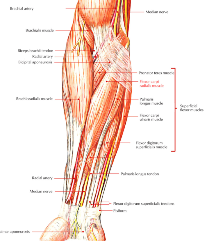
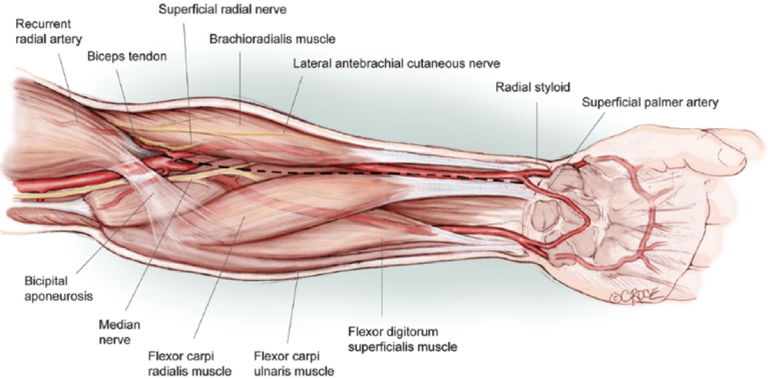
Harvesting the Radial ArteryArie Blitz et al
12.
Oxygen tension in the blood measured by:
Absorbance spectrophotometry
Paramagnetic analysis
Pulse contour analysis
Mass spectroscopy
Paramagnetic Oxygen Analyser
Clarke Electrode
13.
Which phase of the cardiac cycle has the highest sodium flux:
Isovolumetric Contraction
Isovolumetric Relaxation
Ejection
Atrial Systole A)
Isovolumetric ventricular contraction
The beginning of this phase corresponds with the peak of the R wave
This corresponds to Phase 0 (rapid sodium influx) of the ventricular myocyte action potential
The ventricles begin to contract during this period
This contraction increases the ventricular chamber pressure and closes the mitral and tricuspid valves.
As a result, there is a fixed ventricular volume during this contraction
Deranged Physiology
14.
The drug with the lowest clearance:
Vancomycin
Piperacillin-Tazobactam
Clindamycin
Ampicillin
15.
The nerve root of the knee jerk reflex
L1/L2
L3/L4
L4/L5
S1/S2
Just be sure you are answering the correct qn as there are Dermatome, Myotome & Reflex MCQs
16.
The efferent limb of the pupillary light reflex synapses onto post-ganglionic parasympathetic fibres in the:
Olivary Pretectal Nucleus
Edinger Westphal nucleus
Ciliary Ganglion
Contralateral Pretectal Nucleus
This qn was not well recalled but related to the pupillary light reflex – learn the pathways & their connections
The PLR has traditionally been divided into two separate pathways based on the clinical manifestations of the defects in this reflex.
The afferent pathway is composed of both the retinal cells that project to the pretectum as well as their recipient neurons, which project bilaterally to the EW nucleus (Figure 1).
The efferent pathway is composed of the preganglionic pupilloconstriction fibers of the EW nucleus and their postganglionic recipient neurons in the ciliary ganglion, which project to the sphincter muscle of the iris (Figure 1).
When each pretectal nucleus projects bilaterally and synapses in both Edinger-Westphal nuclei (cranial nerve III), the activated Edinger-Westphal nuclei begin the efferent limb of the reflex by generating action potentials. The axons of these preganglionic parasympathetic neurons send the signals along the oculomotor nerve to the post-ganglionic nerve fibers of the ciliary ganglion
17.
The effect of a competitive antagonist on a dose response curve is:
Right shift
Change in slope
Change in peak
Left shift
Deranged Physiology
18.
Drugs with low extraction ratio are primarily affected by
Hepatic blood flow and intrinsic enzyme activity
Hepatic blood flow and plasma protein binding
Protein binding and intrinsic enzyme activity
Hepatic blood flow only
For drugs with low extraction ratio:
only unbound drug penetrates membranes and is available for elimination. An increase of the unbound fraction will thus proportionally increase the clearance
Variation of the organ’s intrinsic ability to eliminate the drug will also slightly affect organ clearance
19.
Which is true regarding the antifungal activity of anidulafungin:
Inhibits glucan synthase
Inhibits ergosterol synthesis
Narrow spectrum limited to yeast organisms
Is affected by renal impairment
Anidulafungin has potent and broad antifungal activity against Candida and Aspergillus spp., including those resistant to fluconazole
It does not undergo metabolism but is eliminated through spontaneous degradation with linear kinetics and a terminal half-life of 40 to 50 hours
Neither end-stage renal impairment nor dialysis substantially alters the pharmacokinetics of anidulafungin.