16A15: Exam Report
Describe the structure and function of the alveolus.
52% of candidates passed this question.
Better answers related structure to function. Many answers lacked key anatomical features (for
example pores of Kohn, basement membrane, interconnecting walls / alveolar interdependence etc.). There was little understanding of the role and origin of the basement membrane of the alveolus. Some candidates went into detailed discussions of Work of
Breathing, respiratory mechanics and the renin-angiotensin system which were not asked for.
Answers not reaching a pass mark generally suffered from lacking detail and suggested only a superficial understanding of the area.
F1i / 16A15: Describe the structure & function of an alveolus
Definition: Alveoli = expanded chambers of epithelial tissue that are the site of gas exchange
- Any surface with an alveolus is a RESPIRATORY ZONE
- Respiratory bronchioles → alveolar ducts → alveolar sacs
- Alveolar ducts = elongated airways surrounded only by alveoli
- Alveolar sacs = spaces surrounded by clusters of alveoli, to which they open into
- ~300 million alveoli = 70m2 (tennis court)
- Each alveolus surrounded by a network of capillaries & elastic fibres
Alveolar Cell Types
Capillary Endothelial Cells
- Continuous from endothelium of circulation
- Connected by loose junctions
- Allows passage of large molecules + MΦs
Alveolar Epithelial Cells – Type I
- Lines 95% of surface
- Joined by tight junction
- Thin = 0.1μm → GAS EXCHANGE
- Nil organelles except for nucleus
- 1 epithelial cell = covered by many capillaries
- No cell division
- Sensitive to damage by ↑[FiO2]
Alveolar Epithelial Cells – Type II
- As numerous as T1 cells, but their shape means they only cover 5% alveolar air surface
- Cuboidal shape (Type 1 thin & flat)
- No role in gas exchange
- Stem cells from which T1 cells arise
- Cytoplasm stores surfactant
- Secretes cytokines
- Resistant to O2 toxicity
Brush Cells
- With short microvilli
- Serve as afferent n. ending receptors
- Monitor air quality
Alveolar Macrophages
- In both connective tissue & alveolar space
- Passes freely from circulation
- Scavenge foreign bodies → bactericidal
Pores of Khon
- Opening in alveolar septa
- Allow circulation of air from one alveolus to another
Collateral Ventilation
→ Pores of Khon
→ Martin’s channels
→ Lambert’s channels
- Collateral ventilation = ventilation of alveoli via pathways that bypass the normal airways
- Presence can be beneficial (↓resistance, allow gas exchange) or harmful (easy extension of disease)
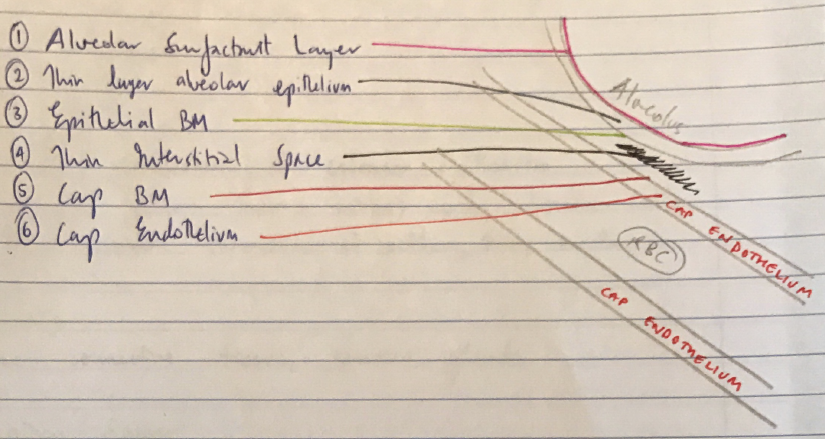
Alveolar – Blood Barrier (aka respiratory membrane)
- The layers which gases must diffuse between:
- Alveolar surfactant layer
- Thin layer alveolar epithelium
- Epithelial BM
- Thin interstitial space
- Capillary BM
- Capillary endothelium