F1ii: Outline the anatomy of the chest wall + Describe its function in respiration
- Thorax = superior part of trunk between neck & abdomen
Fascia
- Skin
- Subcutaneous tissue: fat, sweat glands, blood, lymphatics, cutaneous nerves, breasts & mammary glands (mature females)
- Deep fascia: thin fibrous membrane → inverts underlying muscle & tendons → barrier to infection
Skeleton
Sternum
- Flat, elongated bone at midline of thoracic cage
- 3 parts: manubrium, body, xiphoid process
- 3 indentations: jugular notch, clavicular notches, costal notches
Thoracic Vertebrae
- 12 → long spinous processes
- Costal facets on body → articulation with rib tubercles
- Costal facets on T process → articulation with rib tubercles
Rib
- True ribs: 1 – 7 → Attached directly on sternum with own costal cartilages
- False ribs: 8 – 10 → Their cc attach to rib above them ∴their attachment to sternum is indirect
- Floating rib: 11 – 12 → Attached to vertebrae but not sternum ∴ ‘floating’
Typical Rib
- Posterior portion → costal angle → body
- Posterior portion → 2 facets to articulate with vertebrae
→ Neck
→ Tubercle to articulate with T process of T vertebral body
- Body → long curved shaft of bone
→ anteriorly attaches to cc → sternum
- Costal angle → rib is twisted on its axis to create an angle
→ here where it ∆ direction is its weakest link
Atypical Ribs
- Ribs 1 – 2
- Shorter & flatter
- Rib 1 forms thoracic inlet & manubrium transmitting GREAT VESSELS, OESOPHAGUS, TRACHEA, NERVES + LYMPHATICS
- Ribs 11 – 12
- Short & floating
- No sternal attachment
- ∴ no neck/tubercle
Rib Movement
- Ribs 1 – 7 attach sternum + spine → ∴ limited mobility
- Ribs 8 – 10 have longer CC ∴ ↑mobility
- Ribs 11 – 12 float ∴ greatest mobility
∴ all ribs have different ROM
- Thoracic wall moves like a bucket handle
- It moves laterally + up
- When upper ribs are elevated the A-P diam of thorax increases → movement resembling a pump handle
Muscles of Chest Wall
- Serratus posterior superior → elevate ribs
- Serratus posterior inferior → depresses ribs
- Levator costanum → elevates ribs
- External IC → elevates ribs
- Internal IC → depresses ribs
- Innermost IC → elevate ribs
- Subcostal → elevate ribs
- Transversus thoracic → depresses ribs
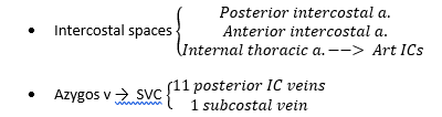
Arteries & Veins (intercostal spaces)
Nerves
- 12 pairs of spinal nerves
- Divide into ventral & dorsal rami to supply IC space, bones, joints & muscles of thoracic wall
Chest Wall Muscles & Respiration
- Respiration movements of chest:
Inspiration
- Requires thorax expansion
- ↑AP chest diameter
- ↑lateral chest diameter
- ↑vertical chest diameter
Passive inspiration: diaphragm + external ICs
Forceful inspiration: Scaleni, SCM, serratus anterior, serratus post superior, pec major + minor, levatores costorum
- Pump bundle of sternum elevates = ↑AP diameter
- Bucket handle ribs elevate = ↑lateral diameter
- Lowering of diaphragm = ↑vertical movement of thorax
- ∴ ↓ intrathoracic P & atmospheric air enters lungs
Expiration
- Passive process, the opposite of inspiration
Quiet expiration: internal ICs
Forceful expiration: Traversus thoracic, rectus abdominis, internal + external obliques, serratus post inf.
- Pump handle of sternum lowers = ↓AP diameter
- Bucket handle elevation of ribs lowers = ↓lateral diameter
- Diaphragm elevates → ↓vertical length thorax
∴ ↑intrathoracic P → air pushed back to atmosphere