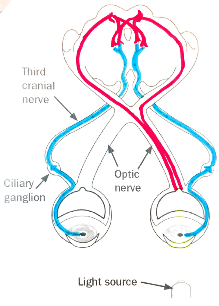
EWN projects pre-ganglionic parasympathetic fibres which travel along CN III & synapse in CILIARY GANGLION onto post-ganglionic parasympathetic fibres
Post ganglionic parasympathetic fibres innervate sphincter m. of pupils à PUPILS CONSTRICT
= Direct & consensual light reflex
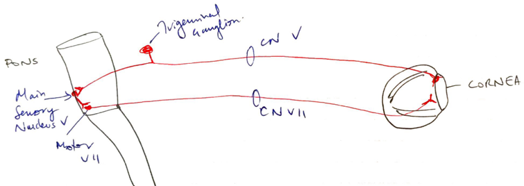
Corneal Reflex
→ CN V TRIGEMINAL N (OPTHALMIC BRANCH) Afferent
→ CN VII FACIAL N Efferent
Light touch receptors in conjunctiva & cornea stimulated
Pass via OPTHALMIC BRANCH of TRIGEMINAL N & through Trigeminal Ganglion to Main Sensory Nucleus of Trigeminal n.
Interneurons transmit to Motor Nuclei of Facial n.
Facial n. supplies ORBICULARIS MUSCLE
Closure of eyelids
Reflex Response to Pain in Trigeminal Distribution
CN V TRIGEMINAL N
CN VII FACIAL N
(Apply pain to supraorbital region (V) → No facial movement (VII))
Main Sensory Nucleus lies in pons & receives light touch sensation
Postganglionically Trigeminal N. divides into OPHTHALMIC, MAXILLARY & MANDIBULAR NERVES
Interneurons transmit to Motor Nuclei of Facial N (in Pons near border with Medulla)
Facial N. carries motor control to all facial m. except eye muscles
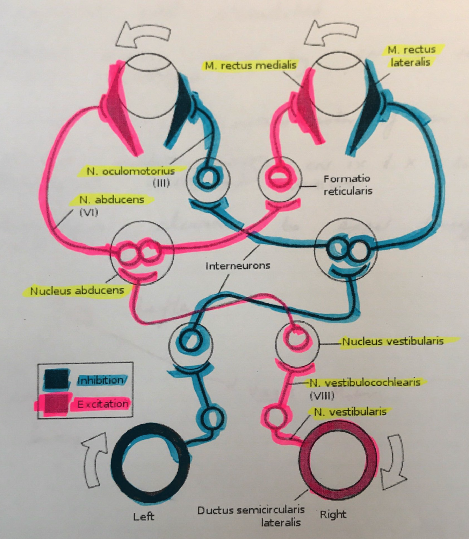
Vestibulo-Ocular Reflex (VOR)
CN III OCULOMOTOR N
CN IV TROCHLEAR N (Vertical response)
CN VI ABDUCENS N (Horizontal response)
CN VIII VESTIBULOCOCHLEAR N
Vor Test Basis
Activation of vestibular movement causes eye movements
Reflex to stabilise visual field on retina
Move head → eyes move opposite direction
VOR does not require visual input & can be elicited by caloric (cold/hot) stimulation of inner ear & in total darkness!
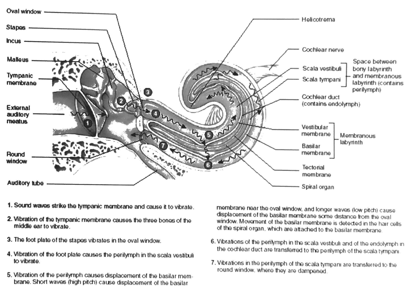
Caloric Testing
Ice cold (<30°C) water is irrigated into external auditory canal
Temperature difference b/w body & water creates a convective current in endolymph of semicircular canal
If water COLD the endolymph falls
→ ↓rate of vestibular firing → mimics head turn to contralateral side
→ eyes turn to ipsilateral side (fast movement) & nystagmus (slow movements) to correct towards contralateral ear
COWS = Cold opposite, warm same
WARM water → endolymph rises → ↑firing vestibular afferent n. → mimics head turn to ipsilateral side → both eyes turn to contralateral ear (fast) & ipsilateral ear (slow = nystagmus) to correct
∴VOR elicits = PHYSIOLOGICAL NYSTAGMUS
Nystagmus = rhythmic abnormal eye movements
Slow movement driving eye off target
Fast follow up movement bringing eye back to target
∴Absence of VOR = Brainstem not working
Vor Test
Cold H2O causes endolymph to fall
↓Vestibular N (CN VIII) afferent firing
Mimics head turn to CONTRALATERAL SIDE of H2O irrigation
Transmitted via Vestibular ganglion to vestibular nucleus in brainstem
Vestibular nuclei fibres cross to Contralateral Abducens Nucleus (CN VI)
In the abducens nuclei, they synapse at 2 pathways:
Direct to IPSILATERAL Lateral Rectus Muscle via Abducens n (VI)
To CONTRALATERAL Medial Rectus Muscle via Oculomotor N (III)
Also inhibitory pathways in IPSILATERAL abducens nucleus so that ipsilat. (to cold stimulus) the lateral rectus & contralateral medius rectus are inhibited
Slow movement eyes towards IPSILAT
Fast movement (NYSTAGMUS) to CONTRALAT
Abnormal HORIZONTAL (III, VI, pons)
Abnormal VERTICAL (III, IV, Midbrain)
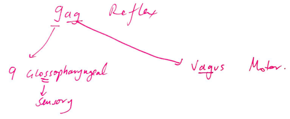
Pharyngeal (GAG) Reflex
CN IX GLOSSOPHARYNGEAL Afferent
CN X VAGUS Efferent
Pharyngeal wall stimulated
Afferent sensory carried by CN IX to Nucleus Ambiguus
Nucleus Ambiguus contains motor of CN IX, X, XI
Efferent output through CN IX & X to pharyngeal constrictor muscles
Pharyngeal elevation to expel foreign object
Cough Reflex
CN X VAGUS Afferent & Efferent
Irritant receptors on posterior wall trachea, pharynx & tracheal carina
Via Internal Laryngeal N. branch of the Superior Laryngeal n. of the Vagus (X) to Medulla
Motor signals transmitted via Superior Laryngeal n. of Vagus (X) to glottis, external IC & diaphragm
Diaphragm (phrenic) & external IC (intercostal n’s) contract
Negative intrathoracic P
Air rushes into lungs
Glottis shuts (Recurrent Laryngeal N. of Vagus X) & vocal cords shut
Abdominal m. & resp m. contract simultaneously generating v. high P > 300mmHg