Wiii: Describe the physical principles of ultrasound and the Doppler Effect
Biological Variable
The attenuation of high frequency sound waves (5 – 15 MHz) at tissue interfaces with variable density
- Sound = a form of mechanical E that travels in a longitudinal wave in a series of compressions (high P) & rarefractions (low P)
- Sound energy is attenuated as it passes through tissue because of:

- ABSORPTION = tissue absorption of sound, responsible for most attenuation of ultrasound wave
- REFRACTION = the change in direction (bends) of a sound wave, occurs when the sound strikes the boundary of two tissues at an oblique angle
Image turns out unclear i.e. ‘2 needles’ traversing a vessel
- REFLECTION = a reflection (echo) is how we identify structures
Reflection occurs when the sound wave contacts tissues with different densities (& part of the beam reflects)
- SCATTERING = when the sound wave is bigger than the object it contacts (i.e. RBC) the wave does not reflect back to the probe, it scatters in all directions
Sensor
- Probe containing a Piezoelectric transducer (PZT)
- Piezoelectric Effect = electrical voltage is applied to quartz crystal & crystal changes dimension
∴ converts Electric E → sound E
- Each PZT has ~250 elements
- Electrodes either side of PZT
- Voltage applied → crystals vibrate → sound emitted
- These crystals spend:
- 1% time emitting sound
- 99% time listening back for sound
- On the way back, use of Reciprocal Piezoelectric Effect calculates how much reflection is occurring
- PROBES all emit at different frequencies
Probe
Frequency
Depth
Linear
Frequency
15 – 6 MHz
Depth
6cm
Curved
Frequency
8 – 3 MHz
Depth
15cm
Cardiac
Frequency
5 – 1 MHz
Depth
35cm
i.e. Higher frequency probes have better resolution but shallow depth
Integrator
- Reflection returns to probe & is transduced by Reciprocal Piezoelectric Effect (Sound E → electrical current)
- Central processor calculates the distance b/w transducer & object according to the speed of sound (1540m/sec) → ∴ uses this to calculate depth
- Measures intensity of a signal → which is displayed as brightness
RESOLUTION = how fine a detail can be seen in an image
- Axial Resolution = the ability to display small targets along beam path as separate entities

- Lateral Resolution = the ability to separate targets perpendicular to the beam path
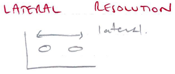
- Temporal Resolution = the ability to show changes in anatomy over time esp imp with echo
Output/Mode
- B – Mode (brightness node)
- 2D cross-section through body
- M – Mode (motion mode)
- Movement of structures over time
- Cardiac scans can be timed with ECG
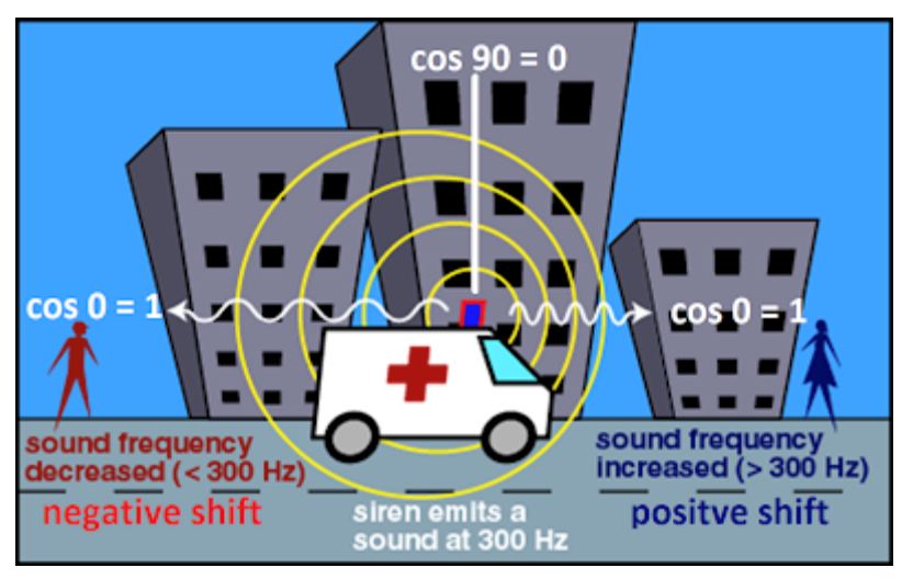
Doppler Effect
- The change in sound pitch as the sound wave changes frequency
Think ∆ pitch as ambulance gets closer/further away from you → Replace yourself with the probe
- Sound waves reflected off objects move away/towards transducer (usually blood)
- Object moving towards transducer → ↑frequency sound wave, +ve Doppler shift
- Object moving away from transducer → ↓frequency sound wave, – ve Doppler shift
- Doppler shifts are calculated by the cosine of the angle
∴ Maximal Doppler shift at 0 degrees (cosine of 0 = 1) when flow is either directly toward/away from transducer