Skip to content
Home
Syllabus
New Syllabus Topics Fourth Edition (2023)
A – Pharmaceutics
B – Pharmacokinetics
C – Pharmacodynamics
D – Variability in Drug Response
E – Cellular Physiology
F – Respiratory System
G – Cardiovascular System
H – Renal System
I – Body Fluids and Electrolytes
J – Acid Base
K – Nervous System – Including Pain
L – Musculoskeletal System
M – Autonomic Nervous System
N – Liver
O – Gastrointestinal System
P – Nutrition & Metabolism
Q – Haematological System
R – Thermoregulation
S – Immunology & Host Defence
T – Microbiology
U – Endocrine System
V – Obstetrics
W – Principles of Measurement and Equipment
X – Procedural Anatomy
Year
Pharmacopeia
MCQs
2025
2024
2023
2022
2021
2020
2019
MPS
Test Yo’Self
Hacks
About
Search
Home
Syllabus
G
G2
G2i: Slow action potential
Home
Syllabus
G
G2
G2i: Slow action potential
Site:
SA Node, AV Node, ventricular conduction system
Duration:
150 milliseconds
RMP:
-60mV
Less negative because their membranes are naturally leaky to Na
+
/Ca
2+
At
-55mV
Fast Na
+
channel n gates already closed ∴inactive
∴only slow type Ca
2+
channel can cause an AP
∴rate of depol. is slow & Ph 0 upstroke is slow with less amplitude → hence name ‘Slow AP’
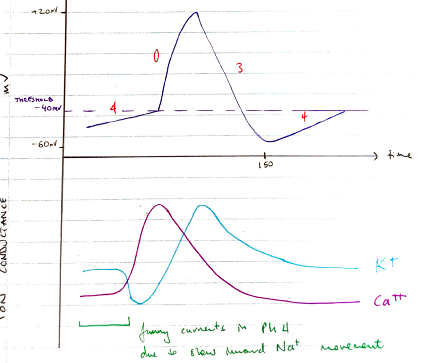
Features
• Ph 0: Depolarisation
Membrane depolarised to
-40mV
Slow L-Type Ca
2+
channel open
Inward Ca
2+
movement
(slow
∴slow upstroke)
Transient ↓K
+
permeability further contributes to depolarisation
• Ph 3: Repolarisation
Delayed rectifier K
+
channel opens
↑K permeability repolarises cell
Ca
2+
channel closing & inactive
Ph 3 ends when membrane potential reaches
-65mV
• Ph 4: Spontaneous Slow Decay
Mechanism unclear
Multiple ionic currents involved
↓K
+
permeability
Slow inward Na
+
movement
KA
‘
funny current’
↑Ca
2+
permeability through T-type Ca
2+
channel
(T = transient)
Then as begins to approach threshold, the L-type Ca
2+
channel opens
NB: CCB do not block the T-type Ca
2+
channels
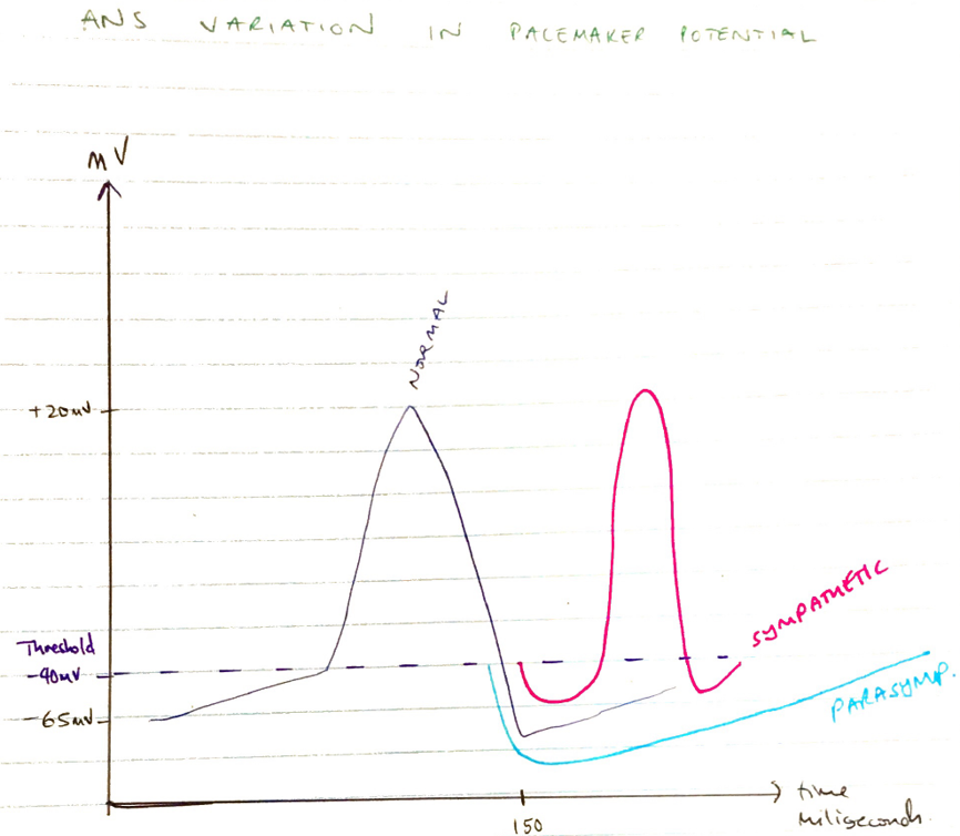
Regulation of Slow AP
Pacemaker potential seen in cells of cardiac excitatory system
Denervated heart → SA Node fires at rate 100bpm
At rest, vagal tone predominates → ∴AP generated ~70bpm
This high intrinsic rhythmicity suppresses automaticity at other loci
“Characteristics of PM potential are predominantly under control of ANS”
ANS influences rate of PM firing
Alters
Slope of Ph 4
Threshold trigger of Ph 0
Degree of hyperpolarisation at end of Ph 3
Parasympathetic
↑K
+
out
& ↓Na
+
& Ca
2+
in
Decrease slope Ph 4 ∴more time taken to reach threshold = ↓discharge rate
More negative Ph 4 membrane potential
Sympathetic
↓K
out
& ↑Na
+
& Ca
2+
in
Increase slope Ph 4 → decrease time to reach threshold → faster discharge rate
Notes on Spontaneous Decay
Not really a ‘RMP’ in PM cell
Constantly decaying ‘MP’
Multiple ionic currents responsible influenced by cAMP
∴SYMP = ↑cAMP = ↑Ca
2+
influx = steeper Ph 4 & rate of d/c
Other factors all affect
time to threshold
(Rate of Decay)
O
2
Thyroid hormones
(↑Ca
2+
& ↑[Adv rec])
Temp
K
Author:
Krisoula Zahariou