G2ii: Outline normal impulse generation in heart and conduction in the heart. Describe the features in a normal heart that prevent generation and conduction of arrhythmias
Electrical conducting system = most important control of cardiac cycle
Impulse Generation
- Occurs in specialised PM cells
- No true RMP
- Spontaneous slow decay
- Their membrane naturally leaky to Na+/Ca2+
- Reaches threshold = -40mV
- Slow Type L Ca2+ open
- Slow inward Ca2+ movement
- Slow upstroke (depol)
- Spontaneous decay influenced by ANS
- SYMP: ↓K out & ↑Na/Ca in
- =↑ slope Ph4
- =↓time to reach threshold
- =Faster d/c rate
- PARASYMP: ↑K out & ↓Na/Ca in
- =↓ slope Ph4
- =More time taken to reach threshold
- =↓d/c rate
- =more negative membrane potential ∴more time taken to reach threshold
- SYMP: ↓K out & ↑Na/Ca in
Normal Conduction Pathway
- SA Node = high intrinsic frequency = 100bpm
- Suppresses automaticity at other loci
- Basal vagal tone → ∴normal HR ~70bpm
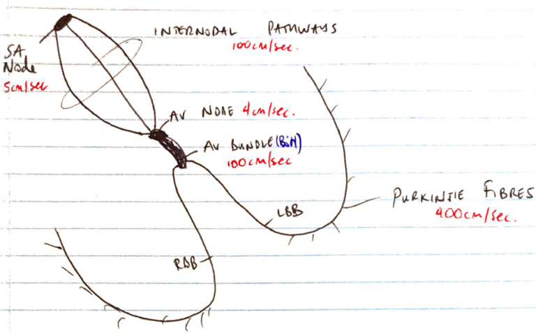
- Impulse travels via 3 internodal pathways (ant., middle, post.) to AV node
- @AV node 13sec delay due to ↓ no. gap junctions
- Travels to Bundle of His
- Down R & L bundle branches
- Divide into PURKINJE FIBRES
- Small fibres project through myocardium
- Rapid propagation of AP @400cm/sec
- Papillary contracts before ventricles → prevents regurg. of blood via AV valves
- IV septum (except base)
- Endocardium
- Epicardium
- Last = Post basal epicardial & basal IV septum
- Almost as soon as signal reaches Purkinje, it’s transmitted to entire ventricle mass for organised ventricular contraction
Features Preventing Abnormal Conduction
- Conducting pathways
- Conducting tissue has multiple IC discs & gap junctions
- If upstream PM is blocked, down stream PM can take over
- Fibrous skeleton & AV node
- Fibrous skeleton prevents conduction between atria & ventricles
- AV node conducts v slowly
- Allows full atrial contraction
- Prevents conduction of rapid rates >220bpm
- Ensures one way conduction
- Refractory period
- The time taken from Ph 0 until next possible depolarisation of a myocyte
- Cells which are not PM cells (have fast AP) have different degrees of refraction depending on no. of Na+ channels which have recovered from inactive state
→ Absolute refractory = no stimulus can excite myocyte
- RRP = supramaximal stimulus will depolarise the cell & cause an AP (not all Na+ channels recovered yet)
