G3iii: Define myocardial contractility & describe dP/dT, ESPVR, Ejection Fraction

Defintion

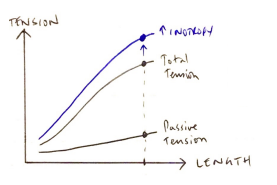
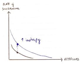
Contractility = the intrinsic ability of myocardial fibres to shorten, independent of preL & afterL

(it augments force of myocyte contraction via SLIDING FILAMENT)
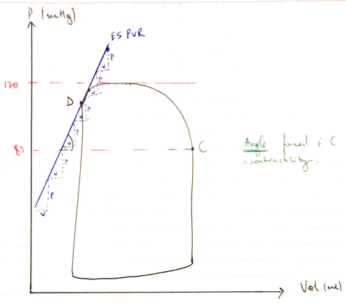
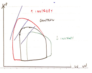
  • ∆ inotropy are caused by cellular mechanisms that regulate the interaction between actin & myosin, independent of ∆ sarcomere length
  • ↑CONTRACTILITY causes:
  1. ↑ Active Tension → so that for any given preL sarcomere length, there is more tension because the inotropy augments the force of the contraction
Tension
Rate
  1. ↑ Rate of Shortening → ↑ inotropy will ↑rate of shortening for any given afterload because of ↑rate cross-bridge turnover e. VMAX

Indices of Contraction

  • Papillary Muscle Force = GOLD STANDARD
  • All other indices depend on HR/preL/afterL
  • dP/dT MAX = maximum rate in ΔP in LV during isovolumetric contraction
    • It is a pre-ejection index independent of afterL (because AoV is shut) but is dependent on preL
    • It is an invasive measurement done in a cardiac cath lab
Indices of Contraction
  • Ejection fraction: the % of EDV ejected from the LV with each contraction
    • Normally >55%
  • EjF affected:
    • Minimally by preL if MV/AoV normal
    • ↓linearly with ↑ afterL
    • Inverse relationship with HR
  • May be measured by Doppler wave assessment of MR jet (which occurs in IVC MV jet velocity ∆ is reflecting the LVP ∆)
  • ESPVR: the maximal P generated by the ventricle at any given LV volume
    • At end of systole the sarcomeres are maximally activated ∴you can measure the P generated in the LV for a given volume
  • On the PV loop
    • Starts just after 0mL
    • Intersects D (AoV shut)
    • Straight line
    • Intersects SBP (120) & DBP (80)
    • Angle of line it makes with C = contractility
Contractility
  • As you ↑/↓ vol, the LVP changes

P/V = Elastance

∴slope of this line is elastance, which is KA E0 → where the PV loop falls on the ESPVR line

  • ↑slope of ESPVR = line moves LEFT
  • ↓slope of ESPVR = line moves RIGHT

Factors Influencing Myocardial Contractility

  • Contractility depends on:
    • Substrate supply
    • Integrity of myofilaments
    • Coordinated electrical depolarisation
    • Metabolic homeostasis
    • Coronary blood flow

↑ C

  • SNS through ↑Ca2+ release

↓ C

  • PNS through ↓Ca2+ release
  • Hypercapnia = ↓pH = ↓Ca2+ release
  • Volatiles = direct – ve inotrope