G6iii: Waveforms & pressures seen in each anatomical location during insertion of a PAC

Definition

PAC = the PAC is a balloon-tipped thermodilution catheter 110cm long, that is inserted via a large vein & floated into the PA
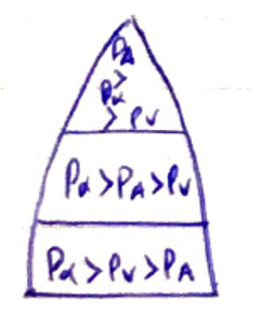
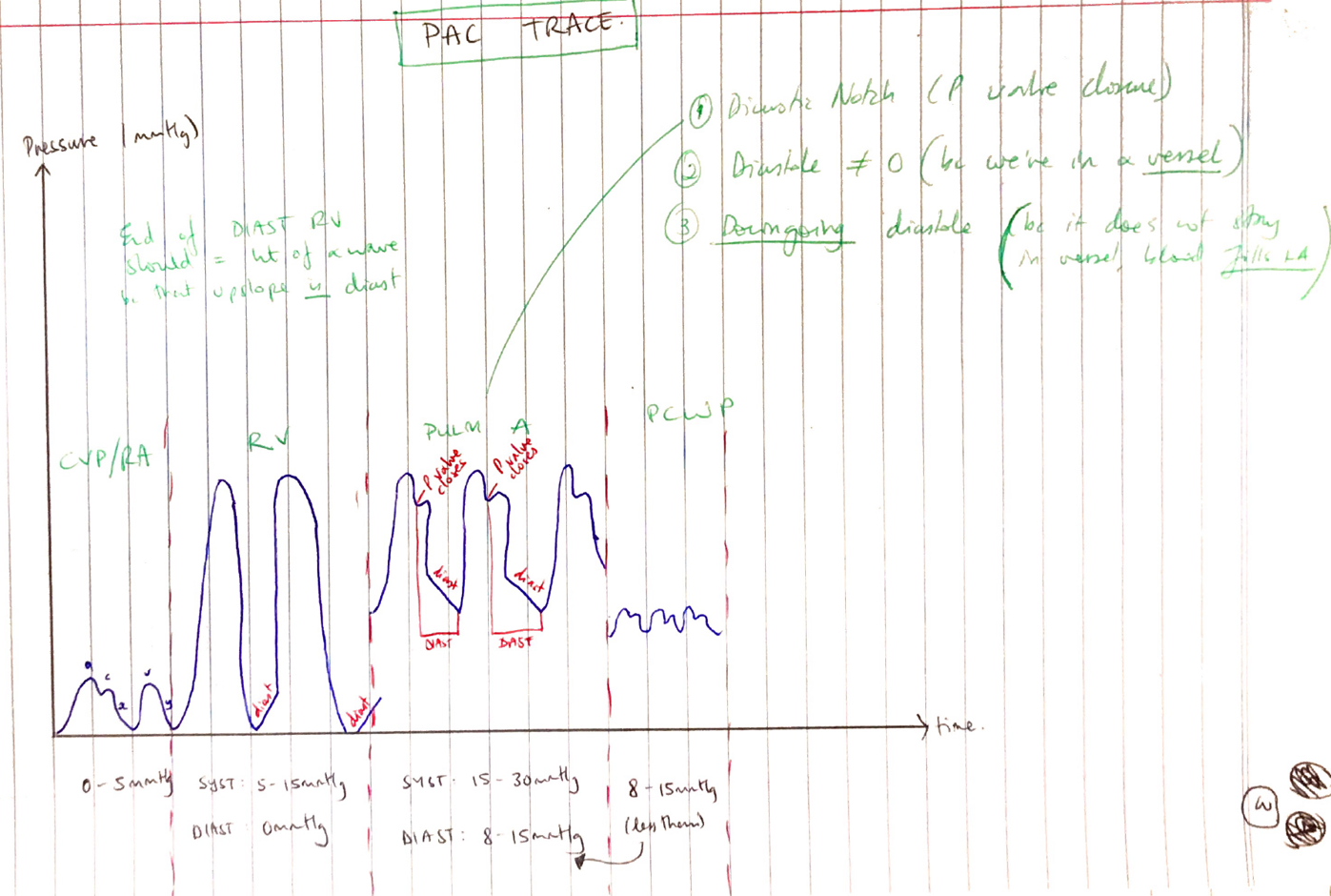
Indications

  1. Measure CO (via thermodilution)
  2. Central temperature monitoring
  3. Measure intracardiac pressures (CVP, RA RV, PA)
  4. Estimates LV preload (via Wedge Pressure)
  5. Sample mixed venous blood

Insertion

  • Sterile Seldinger Technique
  • Through a sheath into central vein
  • Connected to a pressure transducer to ‘map/ your run
  • Once RV waveform seen, balloon inflated to facilitate PAC progress through R) heart
  • Once PA is reached, PCWP/waveform is used to confirm position

CVP/RA

  • Pressure 0 – 5mmHg
  • a wave = atrial contraction
  • c wave = isovolumetric contraction
  • x desc. = beginning of systole where RV pulls TV
  • v wave = venous return (RA filling)
  • y desc. = passive RV filling

RA

  • Systole 15 – 30mmHg/Diastole 0mmHg
  • Upstroke in diastole (filling)
  • Systole without dicrotic notch

PA

  • Systole 15 – 30mmHg/Diastole 8 – 15mmHg
  • Dicrotic notch (pulm. valve closure)
  • Downstroke diastole (because it’s a vessel & fills LA)
  • Diastole ≠ 0 because we’re in a vessel

Pulmonary Wedge

  • Estimates LVEDP & ∴ indicator of LVEDV (LV PreL)
  • 5 – 15mmHg
  • Should look like RA trace but massively overdamped ∴looks like a squiggly line

PCWP

  • Wedging provides a static column of blood between PA & LA
  • Allow estimation of LVEDP (and indicates LVEDV)
  • Must be in WZ3
  • Flow is best in WZ3


  • You don’t want PA influencing PA trace


  • NB: Zone 2 has best V/Q matching
  • By convention measured at end expiration & end diastole (ECP p wave)

Confirming Placement

  1. In WZ3 below RA on XR
  2. Should not vary >50% with PEEP (because then Alveolar P is affecting it too much)
  3. PCWP should be less than diastolic P in PA

Measurements

Direct

RA pressure

RV pressure

PA pressure

PCWP

CO

Mixed venous oxygen saturation

HR

Indirect

CI

SV

SVI

SVR

SVRI

PVR

PVRI

CaO2 – arterial oxygen content

CvO2 – mixed venous oxygen content

DO­2 – oxygen delivery

VO2 – oxygen consumption

  • RV diastolic must be lower than CVP, so RV can fill in diastole
  • PCWP wave similar to CVP but higher
  • PA diastolic must be higher than RV diastolic
    • PA is a vessel, diastole ≠ 0
    • PA diastole is down-going because it is a vessel, blood doesn’t stay there, it fills the LA

Pathology That Alters Valves

CVP/RA

  • Fluid overload
  • RV dysfunction
  • TR/TS
  • Tension/Tamponade
  • Severe LV dysfunction/MV dysfunction

RV

  • Pulmonary hypertension
  • LV dysfunction (bulging LV septum)
  • Tamponade

PAP

  • Any cause of Pulmonary HTN
    • Idiopathic
    • Vasculitis
    • Raised pulm. venous P
    • Chronic lung disease
    • Valvulopathies

PAWP

  • Fluid overload
  • PEEP/↑intrathoracic P
  • Valvulopathies (MV)

Advantages

  • Directly measure & indirectly derive wide range haemodynamic variables
  • Non-toxic “indicator” (temperature)
  • Extra lumens for drug administration
  • Once inserted, can repeat measurements for trend/guide fluid therapy

Disadvantages

  • No study demonstrate I/O PAC = improved clinical outcome
  • Skilled operator
  • Misinterpretation of data
  • Many variables can make data inaccurate

Injection

Slow bolus

Inaccurate vol.

Inaccurate temp.

PAC

Malpositioned

Thermistor wedge

Patient

Resp fluctuations (should be measured at expiration)

TR

HCT

Arrhythmias

  • Complications of catheter insertion → many are critical:
    • Arrhyhthmias
    • Knotting of catheter
    • Balloon rupture
    • PA infarction
    • PA rupture
    • Endocarditis
    • Thromboembolism
    • Air embolism
    • Valve rupture