Describe the baroreceptors & their role in the control of blood pressure.
62% of candidates passed this question.
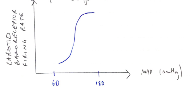
This is a core topic and a detailed knowledge was expected. Baroreceptors are stretch receptors located in the walls of the heart and blood vessels and are important in the short term control of blood pressure. Those in the carotid sinus and aortic arch monitor the arterial circulation. Others, the cardiopulmonary baroreceptors, are located in the walls of the right and left atria, the pulmonary veins and the pulmonary circulation. They are all stimulated by distention and discharge at an increased rate when the pressure in these structures rises. Better answers provided some detail on the innervation for these receptors. It was expected candidates would describe that increased baroreceptor discharge inhibits the tonic discharge of sympathetic nerves and excites the vagal innervation of the heart. This results in vasodilation, venodilation, a drop in blood pressure, bradycardia and a decreased cardiac output.
Some candidates had a major misunderstanding around the purpose of “low pressure baroreceptors” with many believing that these are the ones that respond to lower blood pressures, while the “high pressure baroreceptors” respond to higher blood pressures.
G4iv / 14B16: Describe the baroreceptors & their role in the control of blood pressure
Definition
BP = the pressure in the arterial system = CO x SVR
Important because it allows a pressure gradient to ensure organ blood flow
It is the 1° measured variable in the reflex control of BP
Monitored by BARORECEPTORS
Controlled by a NEGATIVE FEEDBACK SYSTEM
Baroreceptors
Splay type nerve endings
Stretch receptors in the walls (adventia) of blood vessels & heart