F2i / 25B11 / 20A13 / 15B13 / 15A01: Describe the Control of Alveolar Ventilation
25B11: Exam Report
Describe the control of breathing using the following headings:
- sensors (50% of marks)
- controllers (40% of marks)
- effectors (10% of marks)
Include in your answer the location(s) and function(s) of each.
29% of candidates passed this question.
An ideal structure was represented in the breakdown of this question and gave an indication of the level of detail and time candidates should devote to describing the contribution of each to the control of breathing.
This question was generally well answered. Good answers were able to integrate this sensor- controller- effector mechanism including the stimuli and effects.
- Sensors included a detailed description of the central and peripheral chemoreceptors and their relative primary and secondary stimuli. Better answers included other sensors such as pulmonary, skeletal, and baroreceptors and briefly touched on their contribution.
- This section required a brief description of the key areas that control breathing. These can be found in the medullary respiratory centre (CPG, VRG, DRG), the pons and the cortex.
Their functions and primary effects needed to be listed. The role of PaCO2 (major determinant) and PaO2 was also required. - Here only a brief description of the muscles that contribute to inspiration and expiration and the main nervous innervation would have constituted a good answer.
20A13: Exam Report
Explain the control of breathing
53% of candidates passed this question.
Most candidates provided a structured answer based around a sensor / central integration / effector model with appropriate weighting towards the sensor / integration component. Better answers provided an understanding of details of receptor function, roles of the medullary and pontine nuclei and how these are thought to integrate input from sensors.
Marks were awarded to PaCO2 ventilation and PaO2 ventilation response when accurate, correctly labelled diagrams or descriptions were provided.
15B13: Exam Report
Describe the control of alveolar ventilation
42% of candidates passed this question.
The most comprehensive answers were those structured as sensor-controller-effector with an explanation of each part and how homeostasis was maintained.
Insufficient detail was generally provided as to how central and peripheral chemoreceptors were stimulated. A description of central control was required, rather than listing nuclei or areas.
Many failed to address all three components of a control question and focused primarily on the sensors.
Many answers were just too brief and did not present enough information to demonstrate understanding.
15A01: Exam Report
Explain the control of breathing.
71% of candidates passed this question.
This question was generally well done. It was expected answers would include discussion of the three core elements of sensors, a central controller and effectors.
Central control involves three main groups of neurons in the brainstem with some cortical voluntary control also possible.
More in depth answers included graphs of the ventilatory response to oxygen and carbon dioxide tensions.
F2i / 25B11 / 20A13 / 15B13 / 15A01: Describe the Control of Alveolar Ventilation
1° function of lungs is to exchange CO2 & O2
→ to maintain normal levels of PO2 & pCO2 in arterial blood
→ This is kept in tight regulation by the control of ventilation
- 3 elements to Resp Control System
- SENSOR
- CENTRAL CONTROLLER
- EFFECTOR
Central Controller
- Respiratory Centre of Medulla drives the rate & vol of ventilation
- Contains 3 groups of neurons:
- DORSAL GROUP (Insp)
- VENTRAL GROUP (Expr)
- PRE BOTLINGER COMPLEX (Expr)
- Receives sensory input to produce:
- INSP PHASE: gradual ramp ↑of insp n & m activity
- EXPR PHASE I: ↓insp motor n. discharge
- EXPR PHASE II: insp n & m inactive
Effectors
- INSP: diaphragm, external intercostals
- EXPR: internal intercostals, abdominal m’s
Sensors
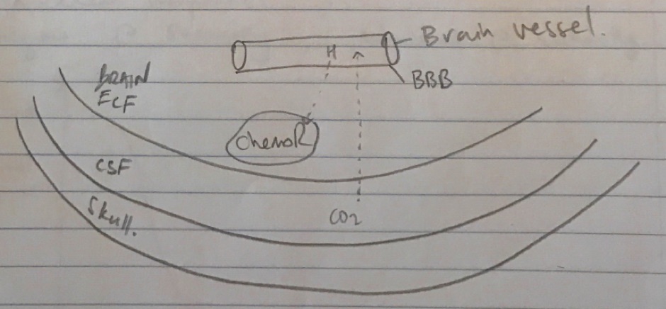
Central chemoR
- Neurons which lie near ventral surface of medulla
- Separate from respiratory centre
- Surrounded by brain ECF (which equilibrates with CSF)
- Stimulated by ↑[H+]
- CO2 of CSF freely diffuses across BBB
- Converted: CO2 + H2O ⇄ H2CO3 ⇄ H+ + HCO3–
- Stimulation of central chemoR → ↑their afferent output → ↑activity Resp Centre → ↑resp muscles
- ∴↑CO2 = ↑H+ = ↑MV
- Central ChemoR v. sensitive to ↑pCO2
- CSF has pH 7.32
- CSF has less buffering capacity than blood (↓[protein])
Peripheral chemoR
- Located @ 2 locations
→ Carotid A. bifurcation → ka CAROTID BODIES → Transmit via Glossopharyngeal (IX)
→ Aortic arch → ka AORTIC BODIES → Transmit via Vagus (X)
- 2 types of cells: T1 & T2 glomus cells
- Carotid body more important cf. aortic bodies
- Respond to pO2, pCO2 & pH
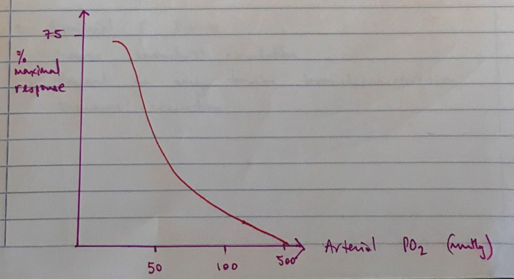
- Main job : respond to arterial pO2
- Respond to O2 tension, NOT Content e. not anaemia, CO poisoning
- Response is RAPID
- ↓pO2 → ↓ATP production & ↑NA, Dopamine, ACh release from glomus cells → ↑afferent discharge → ↑MV
- Non-linear response
- Begins @ 500mmHg, maximal @ <50mmHg
Response to pCO2
- 5% response to pCO2 (majority from central chemoR)
- Rapid responders
Response to pH
- Only by carotid bodies
Cerebral Cortex
- Limbic system & hypothalamus input to Resp Centre
- Emotion, pain, exercise all alter ventilation
- Hyperventilate & breath hold up to a point
Lung receptors
- Stretch receptors
- Located on airway wall
- Respond to ↑lung vol
- Responsible for Hering Breuer Reflex: lung inflation → cessation of breathing → extended expiration
J-Receptors
- Nociceptors in alveolar walls, close to caps of circulation
- Respond to bronchospasm, apnoea, brady, ↓BP
Bronchial C fibres
- Sense bronchial circulation
- Respond to any tension, bronchoconstriction, mucous secretion
Joint/muscle receptors
- Impulses from limb → ↑MV pre-emptively
Baroreceptors
- ↓BP = ↑MV
- ↑BP = ↓MV
Hormones
- NA & Adrenaline = ↑MV
Pain & temp
- Pain, ↑body temp = ↑MV
- Author: Krisoula Zahariou