M1i / 20B05 / 15A20: Anatomy of Sympathetic Nervous System
20B05: Exam Report
Describe the anatomy (70% marks) and effects (30% marks) of the sympathetic nervous system
51% of candidates passed this question.
Most candidates had a suitable structure to their answers, those without a clear organisation of thought tended to gain fewer marks. In many cases incorrect information or limited detail, particularly around the anatomical organisation prevented higher marks.
15A20: Exam Report
Describe the anatomy of the sympathetic nervous system.
25% of candidates passed this question.
A definition of the sympathetic system, followed by a systematic description of the central sympathetic centres; what happens at the spinal cord; the anatomy of the pre and post ganglionic fibres would have been awarded with a pass mark. Additional information about the sympathetic ganglia and the neurotransmitters involved would have rounded off a good answer.
Many answers lacked anatomical detail and described the actions (function) of the sympathetic system which was not asked for.
Most answers lacked any structure. The most common reason for not passing this question was that significant sections of the anatomy from central to peripheral were not mentioned. Most had a simple sketch understanding of the question asked but could not add enough of the next layer to be awarded a pass mark.
M1i / 20B05 / 15A20: Describe the anatomy of the sympathetic nervous system
Definition:
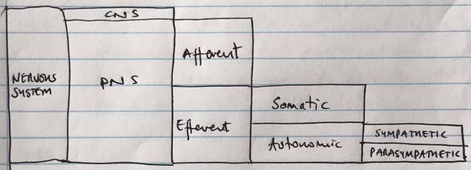
- SNS is a division of the ANS which is activated in fight-or-flight
- ANS is the efferent pathway controlling the action of involuntary organs & tissues in order to maintain homeostasis.

Sympathetic Anatomy
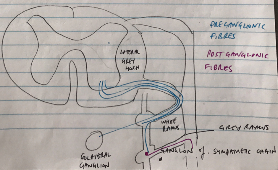
Preganglionic Neurons
- Myelinated slow conducting B fibres
- Cell bodies in intermediolateral horn
- Arise from T1 – L2
- Preganglionic fibre leaves spinal cord via ventral root
- Passes via white ramus into sympathetic chain
- Sympathetic chain = 22 pairs of ganglia
- From sympathetic chain:
- Synapse with post-ganglionic cell bodies at level they entered
- Pass up/down to another level of symp. chain and synapse with that post-ganglionic cell body
- Pass through the symp. chain & synapse with a collateral ganglion
Postganglionic Fibres
- Unmyelinated C fibres
- Either:
- Runs along blood vessels to supply head, neck, thorax & viscera
- Re-enters spinal n’s via grey rami to supply vessels, sweat glands, piloerector m.
- From collateral ganglia synapse close to viscera or in adrenal medulla
NOTE: adrenal medulla is essentially a symp. ganglion in which postganglionic cells have lost their axons & secetes adrenaline, DA, NA directly into the bloodstream
Sympathetic Neurons System Function
The ANS controls the visceral function of the body & is under involuntary control.
The SNS maintains homeostasis during stress.

- Sympathetic neurons originate from preganglionic neurons in grey matter of spinal cord
- From T1 – L2
- Exists spinal cord by ventral roots
- Leave spinal nerve as white rami
- Synapse with postganglionic neuron
- Postganglionic fibres leave as grey rami & join spinal/visceral nerves to innervate target organ
- Sympathetic chain divides into 4 segments:
Cervical (3 ganglia) → head, neck, thorax
Thoracic (T1 – T5) → aortic, cardiac, pulmonary plexi
(T5 – T9) → greater & lesser splanchnic n., renal plexi
Lumbar → coeliac plexus
Pelvic → pelvic plexus
Characteristic
Sympathetic
Origin
Thoracolumbar
Fibre length
Short preganglionic, long postganglionic
Preganglionic fibre
Myelinated B fibres
Ganglia location
Close to CNS
Preganglionic NT
Ach
Ganglia receptors
Nicotinic
Postganglionic fibre
Unmyelinated C fibre
Postganglionic NT
NA (most), ACh (some)
Postganglionic receptors
Adrenergic or muscarinic
Function
Prepares for stress & intense muscle activity
Organ
Action
Receptor
Heart
+ inotropy
+ chronotropy
+ chonotropy
β1
β1 + β2
Arteries
VC
VD
α1
β2
Veins
VC
VD
α1
β2
Lung
Bronchial smooth m. relaxation
β2
GI
↓motility
Sphincter contraction
β2
α
Liver
Glycogenolysis
Gluconeogenesis
Lipolysis
β2 + α
Kidney
Renin release
β1
Bladder
Detrusor relaxation
Sphincter contraction
Β2
α
Uterus
Relaxation
Contraction
Β2
α1
Eye
Pupil dilation
Ciliary relaxation
α1
Β2
- Author: Krisoula Zahariou