F1i / 16B24: Strucure of Trachea Main Bronchi

16B24: Exam Report​

Outline the tracheal (60% of marks) and left and right main bronchial anatomy (40% of marks) in an adult.

14% of candidates passed this question.

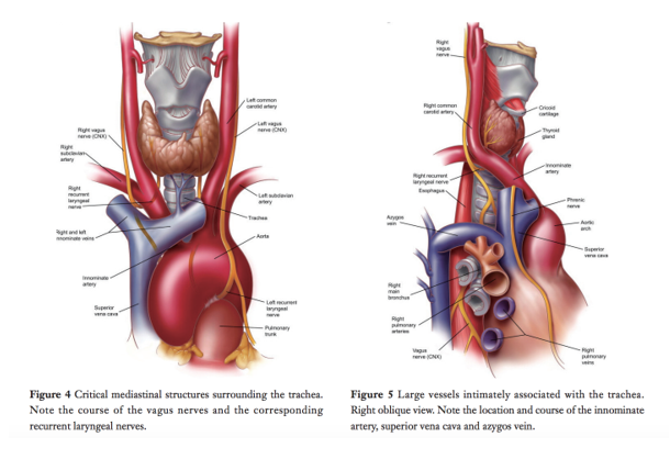
To pass this question, the following were required for each section (trachea and main bronchi): landmarks; basic structural anatomy; and important relations (major vessels; major nerves; major structures).

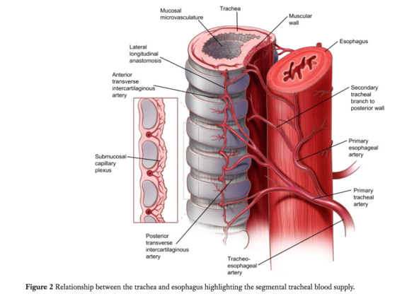
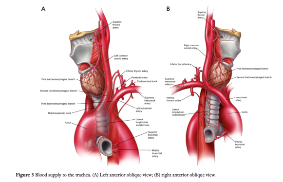
Marks were also allocated for innervation, and blood supply and venous drainage of the trachea.

Most unsuccessful answers did not address a number of these areas. Overall, the answers were better for tracheal anatomy compared to bronchial anatomy.

A structured approach to anatomy questions works well and this was again the case (i.e. relations / blood supply / etc.

F1i / 16B24: Outline the tracheal (60 marks) and left and right main bronchial anatomy (40 marks) in an adult

Trachea & Main Bronchi = Extrapulmonary conducting airways of the tracheobronchial tree

Trachea

R+L Bronchi

Structure

Trachea

Cricoid -> Main Bronchi

11cm long

Anterior: 22 C shaped cartilaginous rings

Post:  trachealis muscle

R+L Bronchi

R+L Main bronchi branch off trachea at T5 (R) and T6 (L)

R main = 3 lobar

*R main is shorter, wider, more vertical

L main = 2 lobar

FN

Trachea

Conducting airway

R+L Bronchi

Conducting airway → supply specific lobes of lung

Layers

Differences

Trachea

Mucosa =

cilitated, pseudostratified columnar epithelium w

goblet (mucous cells),

brush cells (air quality) and neuroendocrine cells

Submucosa = dense connective tissue, mucous glands

Cartilaginous layer – Hyaline C shaped cartilage

Adventia – connective tissue

R+L Bronchi

Mucosa = cilitated, pseudostratified columnar epithelium w goblet (mucous cells), brush cells (air quality) and neuroendocrine cells

Muscularis = continuous layer of smooth muscle that dilates/constricts independent of lung volume

Submucosa = dense connective tissue, mucous glands

Cartilage layer – supporting plates that become smaller distally

Adventia – conn tissue

Major Structures (Fig 4 &5)-

 (Fig 4 &5)-

Trachea

R+L lobes thyroid – anterior to cervical trachea

sthmus of thyroid crosses tracheal ring 3

Oesph – L posterior border of trachea

Vertb bodies – R posterior border

RIGHT Recurrent laryngeal n – dives under R subclavian where it recurs and ascends up to cricoid cartilage

LEFT Recurrent laryngeal n – dives under arch and lateral to ligamentum arteriosum where it recurs and ascends up to cricoid cartilage

Brachiocephalic (1st branch aortic arch) runs L->R over anterolat surface of trachea just under thyroid

L Common Carotid (next arch branch) comes off arch midline to trachea and runs up lateral L border

SVC runs R border trachea to RA

Azygous V runs along R lateral vertb column and joins SVC superior to R tracheobronchial angle

Pulmonary Trunk – anterior to carina – branches with bronchi and runs arterial vessels anterior to bronchi corresponding

R+L Bronchi

L main – crosses ant to oesph

Arterial

(Fig 2&3)

Trachea

Upper Trachea:

Inferior thyroid arteries

Give off tracheoesophageal branches

Lower Trachea:

Bronchial arteries direct from aorta

Arteries of the trachea enter laterally and branch sup&inf in longitudinal fashion (Fig 2)

R+L Bronchi

Bronchial arteries (sup, middle and inf) arise from Aorta

Venous

Trachea

Inf thyroid veins

R+L Bronchi

Bronchial veins

Nervous

Trachea

Trachealis muscle – Symp & Parasymp fibres

Pain – Vagus (X)

R+L Bronchi

Vagus (X)