Xii / 17A13: Anatomy of dorsalis pedis

17A13: Exam Report

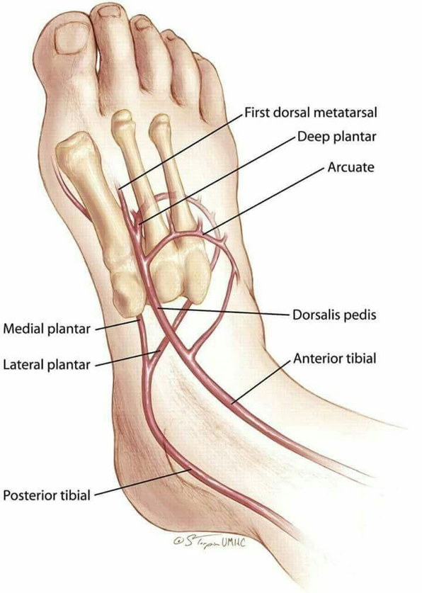
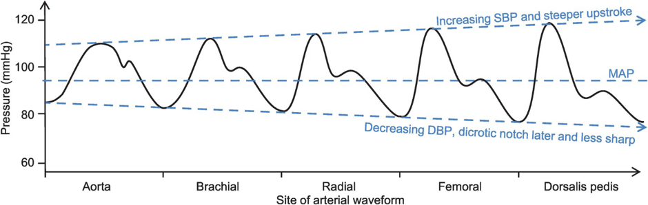
Outline the anatomy relevant to the insertion of a Dorsalis Pedis arterial cannula (50% of marks). Explain the differences between blood pressure measurement at this site compared to measurement at the aortic arch (50% of marks).

30% of candidates passed this question.

The anatomy component of answers frequently lacked required detail. Many candidates listed the observed differences in the waveforms however an explanation for these differences was required.

17A13 / Xii: Anatomy of dorsalis pedis

  • From: Anterior tibial a.
  • To: Proximal 1st metatarsal → divides into the FIRST dorsal metatarsal a. & deep plantar a.
  • Course:
    • Runs with deep peroneal n. (chance of nerve damage)
    • B/w extensor hallucis longus & extensor digitorum longus
    • Crosses ankle joint
  • Lateral: Extensor hallucis longus
  • Medial: Extensor digitorum longus
Dorsal

Aortic Waveform

Due to ejection of blood from LV → Aorta in Systole

Systole

  • Follows R wave on ECG
  • Steep upstroke when AoV opens
  • Reaches peak = SBP
  • Declines
  • Dicrotic notch = closure of AoV

Diastole

  • Follows T wave on ECG
  • Gradual ↓pressure ‘arterial run off’ in diastole
  • Due to Windkessel Effect → Potential E stored in Aortic Elastic Wall in systole converted to Kinetic E that promotes blood flow in diastole

DP Waveform

With increasing distance from aorta;

  • Increasing SBP & Steeper upstroke
  • Decreasing DBP
  • Later Dicrotic Notch
  • Less indentation
DP Waveform