Describe the anatomy relevant to the insertion of an intercostal catheter.
56% of candidates passed this question.
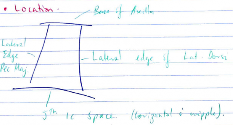
An anatomy question expects the use of anatomical nomenclature to describe relationships. Good answers defined the “safe triangle” for the lateral approach, soft-tissue layers passed through from skin to pleura and relationship of the neurovascular bundle to the ribs and intercostal muscles. Additional marks were gained for describing the anterior approach and related structures. Common omissions included description of deeper structures (relations) including intrathoracic and intra-abdominal organs and level of the diaphragm with regard to rib space. No marks were awarded for a description of intercostal catheter insertion.
Xiii / 18B11: Describe the relevant anatomy for insertion of an INTERCOSTAL CATHETER. How does breathing 100% FiO2 help in the resolution of PTX?
Intercostal catheter = tube inserted into the pleural space to allow drainage of contents
Anatomy
“Safe triangle” → insertion made in the 4th/5th IC space
Layers from skin → parietal pleura:
Skin
Fascia
External IC m.
Internal IC m.
Innermost IC m.
Parietal fascia of thorax
Parietal pleura
Muscle of IC space innervated by neurovascular bundle → sits in groove of rib above
VAN from sup. → inf.
Bundle lies b/w innermost and internal IC m.
100% Fio2 for Resolution of PTX
Adult studies suggest 100% FiO2 improves rate of resolution of PTX
Mechanism:
PTX bubble composed of air: 21% O2, 78% N2, 0.03% CO2, 0.9% Argon, 0.17% other gases
Inspiring 100% FiO2 allows nitrogen washout of PTX bubble
↑PAO2 causes PAN2
N2 moves down concentration gradient from pleural space to alveolus
↓Air in pleural space
Therefore: ↓volume of air in pleural space → overall accelerating resolution of PTX