Outline the functional anatomy of the kidney (40% of marks). Outline the regulation of renal blood flow (60% of marks).
71% of candidates passed this question.
It was expected that answers include sections on the blood supply, the nephron (including the difference between the cortical and juxta-medullary nephrons) and innervation. A number of candidates failed to quantify renal blood flow and to define autoregulation. The concept that it’s the flow that’s regulated was not described by some. Tubuloglomerular feedback was generally described correctly but a reasonable number had the blood flow increasing when an increased sodium was sensed at the macula densa.
17A18: Exam Report
Outline the functional anatomy of the kidneys (40% of marks). Outline the regulation of renal blood flow (60% of marks).
67% of candidates passed this question.
Candidates who scored well weighted their answers according to the marks allocation outlined in the question and adopted a good structure. A number of andidates confused the roles of tubuloglomerular feedback and the renin angiotensin aldosterone pathway.
H1i / 19A04 / 17A18: Outline the functional anatomy of the kidneys (40 marks) & Outline the regulation of renal blood flow (60 marks)
The functional unit of the kidney is the NEPHRON
The nephron is made up of the renal tubule & the renal corpuscle
Each kidney has ~1 million nephrons
A nephron is a hollow tube made of a single cell layer & consists of:
Renal Corpuscle
Proximal Tubule
Loop of Henle
Collecting duct system
Each part of the nephron is made of cells specific to the function of that portion of the nephron → hence, we can describe the functional anatomy
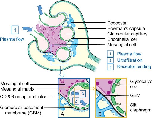
Renal Corpuscle
ULTRAFILTRATE = passive movement of protein free fluid from glomerular capillaries to Bowman’s capsule
Corpuscle is made up of GLOMERULAR CAPILLARIES invaginated by the BOWMAN’S CAPSULE, which is a blind end of the renal tubule
Glomerulus is a capillary network supplied by the AFFERENT CAPILLARY & drained by the EFFERENT CAPILLARY
Fluid passes from Glom Caps → Bowman’s Space by the action of opposing HYDROSTATIC & ONCOTIC PRESSURES
The filtration barrier of the RENAL CORPUSCLE is formed by 3 layers:
Capillary Endothelium
BM
Bowman’s Corpuscle Epithelium
Capillary Endothelium
Fenestrated → i.e. contains holes
Freely permeable to H2O, small solutes, most proteins
NOT permeable RBC, WBC, platelets
Have negatively charged glycoproteins, ∴retards filtration of large anionic proteins
SECRETES FROM BLOOD: H+, NH4+(ammonium), urate, organic ions + cations
Loops of Henle
Thin limbs have basic apical & basolat. membrane
LoH creates an ↑interstitial osmotic gradient in medulla to allow reabsorption of H2O from collecting ducts & production of concentrated urine (1400mOsm/kg) with ADH
Thick Asc. Limb cells have many mitochondria & extensive folding of BASOLAT MEMBRANE
Collecting Duct → 2 cell types
Principle Cells
NaCl reabsorption
K+ secretion
Stimulated by aldosterone
Intercalated Cells
Acid-base balance
*** NOTE: All cells in the nephron except intercalated cells have SINGLE NON-MOTILE CILIA
Mechanosensors: ∆ flow rate
Chemosensors
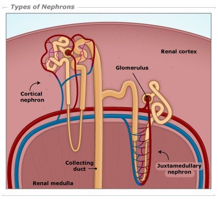
Superficial & Deep Nephrons
Position of corpuscle in cortex determines length of LoH
Superficial cortical nephrons → short LoH
Juxtamedullary (deep) nephrons have their corpuscle close to medulla
They have long loops
Their efferent arteriole forms the PERITUBULAR CAPS & also the VASA RECTA
Peritubular caps = caps around PCT & DCT that function in reabsorption
Vasa recta = caps enveloping LoH that function to maintain [ ] gradient
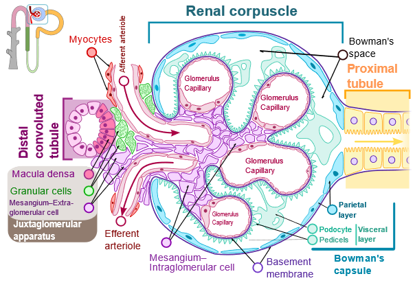
Juxtaglomerular Apparatus
One component of the TUBULOGLOMERULAR FEEDBACK
Juxtaglomerular app. is made of 3 structures:
Macula densa
Extraglomerular mesangial cells
Granular cells
Juxtaglom app. represent distinct part of nephron where Thick Asc. LoH passes through aff. & eff. arteriole
Granular Cells
Of Aff. arteriole
Make & store RENIN
Macula Densa
Of Thick Asc. Limb
In contact with Mesangial cells & Granular cells
Sense passing fluid flow rate
Produce a local VC substance
Extramedullary Mesangial Cells
Of the glomerulus
Contain myofilaments
Contract in response to stimuli
Regulating glomerular filtration
Anatomy
RBF = 20% CO
= 1.2L / min
= 600mL / kidney / minute
RBF is not evenly distributed
90% → cortex
→ flow-dependent function
→ glomerular filtration & reabsorption
→ 5 mL / g / min
→ PaO2 = 50mmHg
10% → medulla
→ outer medulla = 1 mL / g / min
→ inner medulla = 0.2 mL / g/ min
→ Urine concentration
→ PaO2 = 8 – 15mmHg
Therefore, even though medulla is more metabolically active (& extremely vulnerable to hypoxic injury), majority of BF to cortex is to drive filtration of plasma & provide adequate GFR.
RBF Regulation
Q = blood flow
∆P = MAP – venous pressure for that organ
R = resistance to flow through that organ
GFR is strongly influenced by renal perfusion pressures
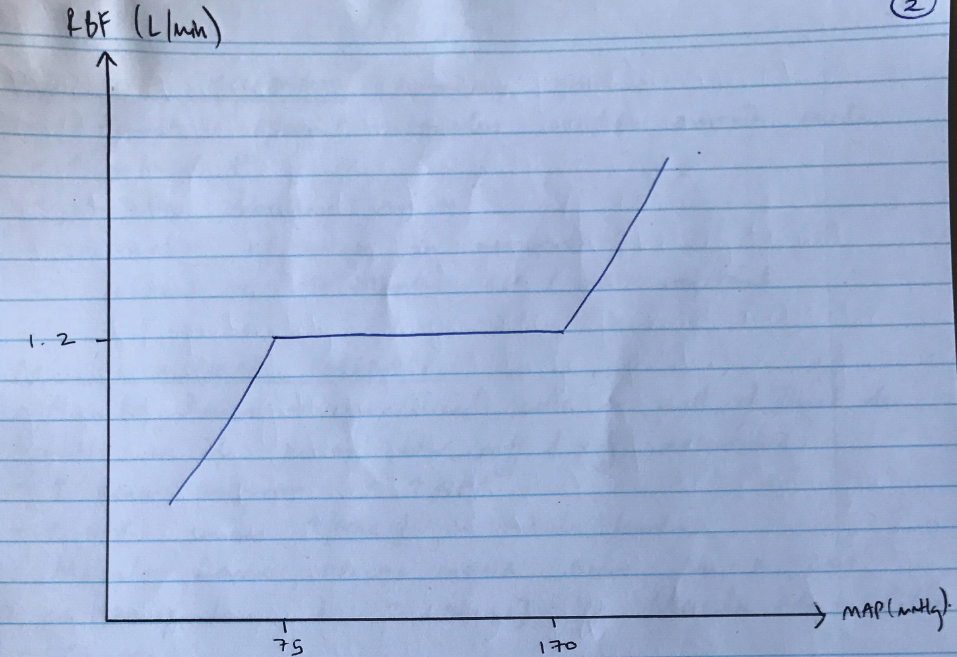
Because of the many activities which can change arterial pressure, the kidneys autoregulate their BF to ensure a constant RBF → & ∴constant GFR over a wide range of pressures
This is KAAUTOREGULATION = the intrinsic ability of an organ to maintain a constant blood flow despite changes in perfusion pressure
Between MAP 75 – 170mmHg, RBF remains constant
GFR remains constant 125mL/min between these pressure as well
R = ∆P/R
∴when ∆P, autoregulation is achieved by changing R (vascularresistance)
Main resistance vessels of kidney are afferent & efferent arterioles
2 mechanisms:
MYOGENIC → responds to ∆ arterial P
TUBULOGLOMERULAR FEEDBACK → respond to ∆[NaCl] of tubule fluid
1) Myogenic Mechanism
Especially quick!
Afferent & efferent arterioles contain smooth m.
↑arterial P
Stretches vascular smooth m.
Contraction of smooth m. in response
∴afferent VC → maintain RBF & GFR constant
2) Tubuloglomerular Feedback
Macula Densa → specialised cells in wall of Thick Asc. LoH as it passes b/w afferent & efferent arterioles
↑renal perfusion = ↑GFR
↑GFR causes ↑[NaCl] in tubule lumen
Macula densa APICAL MEMBRANE have Na-K-2Cl co-transporter
↑[NaCl] in Macula Densa Cells
Causes:
↑ATP release in BLM of macula densa cells
↑adenosine production
Adenosine binds A1 receptors of afferent smooth muscle cells → VC → restores GFR
NB: Macula Densa produce NO if ↓GFR to restore GFR