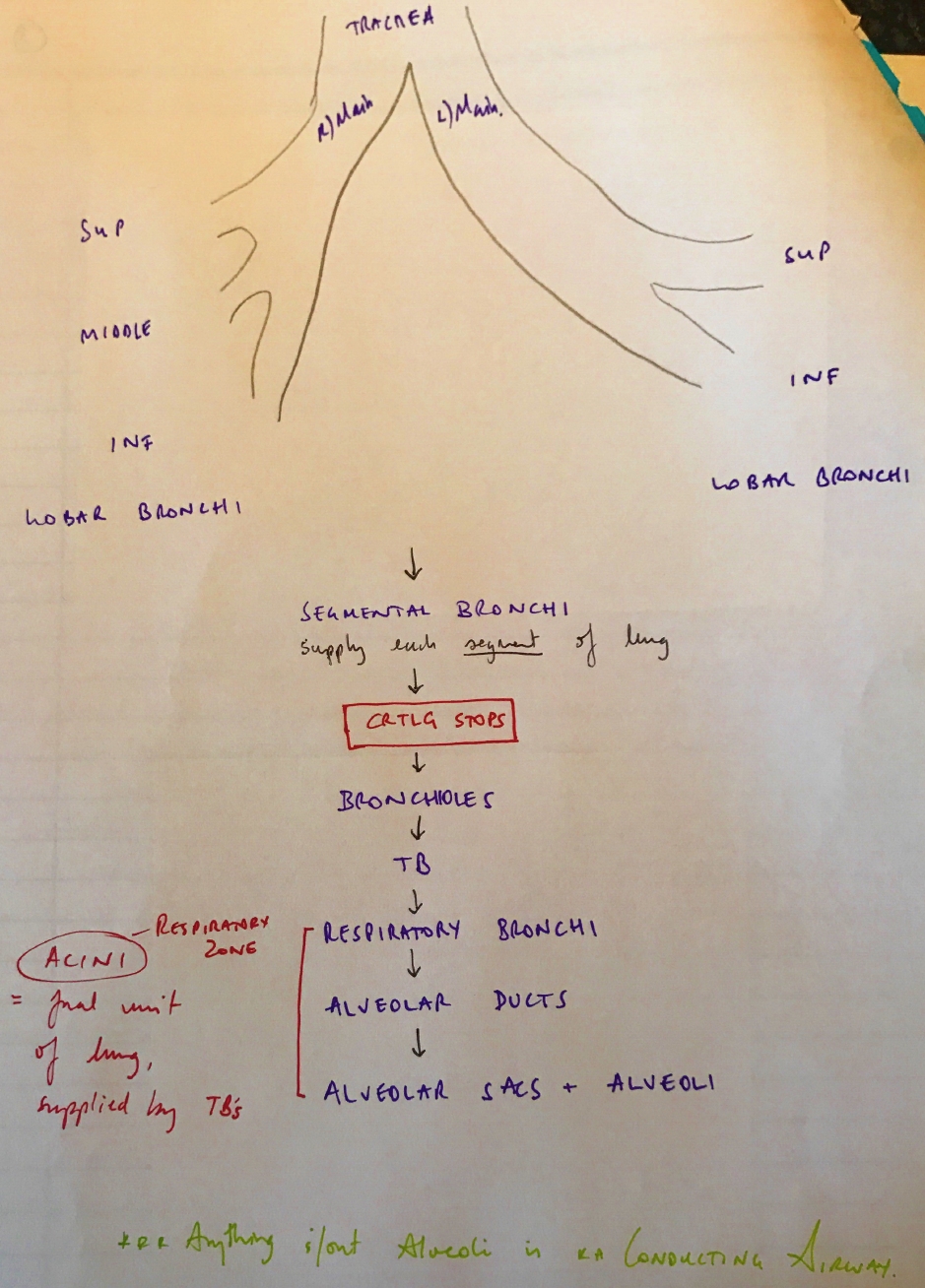
Describe the anatomical course and relations of the trachea and bronchial tree (to the level of the segmental bronchi).
24% of candidates passed this question.
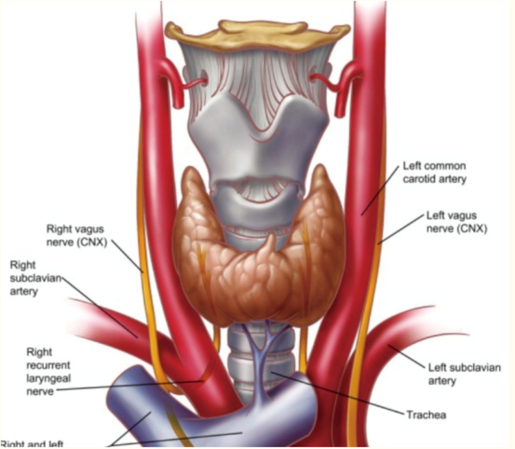
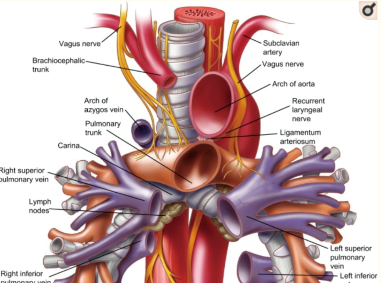
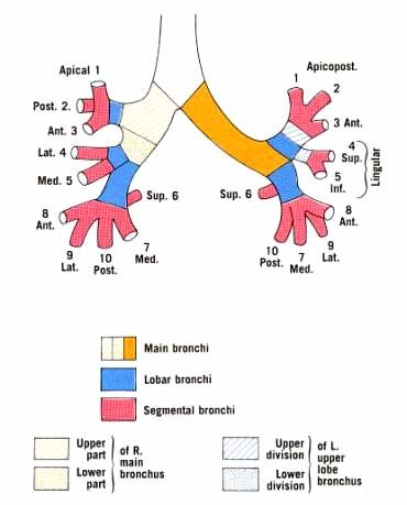
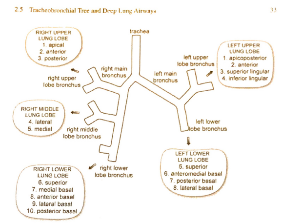
Better answers included details of the significant structures related to the cervical and mediastinal trachea and bronchi. The lobar branches and bronchopulmonary segments requiring naming to attract full marks. Many answers lacked sufficient detail or contained inaccuracies regarding vertebral levels and key structural relations. Some candidates discussed the general anatomy of the airway, including the larynx, structure of the airways, blood supply and innervation. This did not attract marks.
F1i / 19B05: Describe the anatomical course and relations of the trachea and bronchial tree (to the level of segmental bronchi)
Trachea
Structure:
Extends from Cricoid Cartilage → 1° Bronchi
11cm long (extends 15cm w inspiration owing to elastic fibres)