Describe the physiology of skeletal muscle cell contraction.
34% of candidates passed this question.
This question required a description of excitation B contraction coupling. Marks were gained for a brief outline of the structure of a sarcomere and how it facilitates shortening. An explanation of membrane processes, receptor interactions and the contraction processes was required. Mention of the role of ATP was also required and marks were gained for commenting on the mechanism of return to the relaxed state.
Most candidates wrote extensively on the nerve action potential and neuromuscular junction transmission, with minimal reference to events occurring within the skeletal muscle cell membrane. They could not gain marks for this. Few candidates demonstrated knowledge of the ATP dependent walk along processes of myosin heads during contraction.
L1iii / 14A08: Describe the physiology of skeletal muscle contraction
Definitions
Skeletal muscle: Striated muscle tissue which has voluntary control by the somatic nervous system
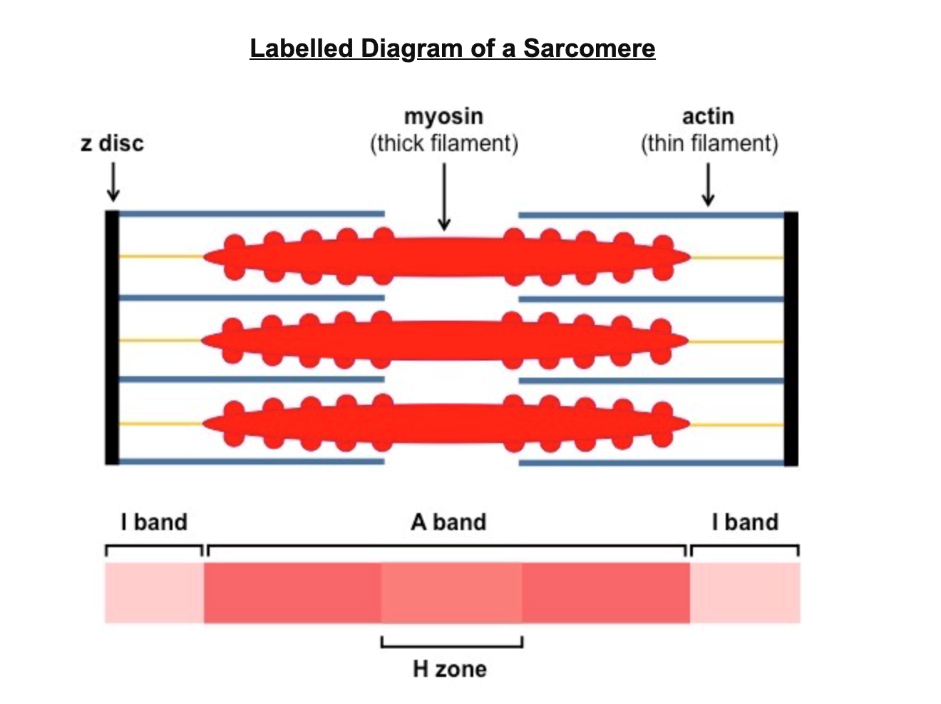
Sarcomere: The structural unit of a myofibril in striated muscle
Excitation-Contraction-Coupling (E-C-C): the rapid events occurring converting an electrical stimulus in the plasma membrane into a mechanical response of muscle contraction
Sarcomere Structure
Macroscopic
Motor unit = nerve ending + muscle fibre
10-100mm diam fibre
Striated, ordered sarcomeres (functional unit)
Microscopic
Multiple parallel myofibrils
Multi nucleated
Mitochondria (aerobic & anaerobic metabolism)
SR (Ca++ storage)
Glycogen (E storage) Myoglobin (O2 storage)
T-tubules
Molecular
Contractile proteins = actin & myosin → Striated
Regulatory proteins = Tropomyosin & troponin
Troponin: protein w 3 subunits
I – inhibits myosin ATPase
C- binds Ca++
T- binds Tropomysin
Tropomysin – blocks actin/myosin interaction
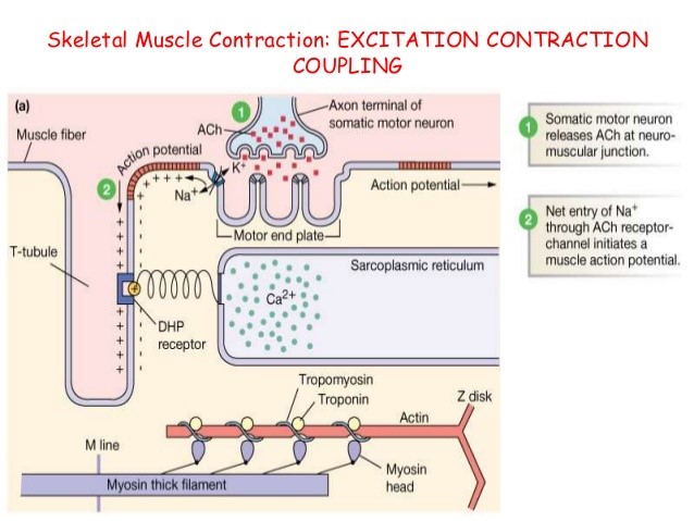
E-C-C
Skeletal Muscle RMP
-70mV
Source of Activation
NMJ
Spread
Via T tubules
AP
Opens fast Na ch
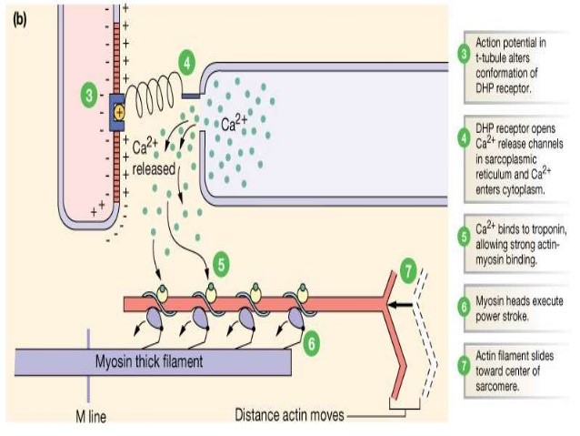
Ca Release
AP propagates along sarcolemma & down T-tubules
Activates L-type Ca++ ch of
T-tubles
Flux of Ca++ intracellularly↑↑↑
Ryanodine Rec of SR open = mass x60↑ Ca++
Cross Bridge
Ca++ binds TnC
Removes inhibition of Troponin-Tropomyosin Complex
Exposes Actin binding site to myosin → commencement of cross-bridging
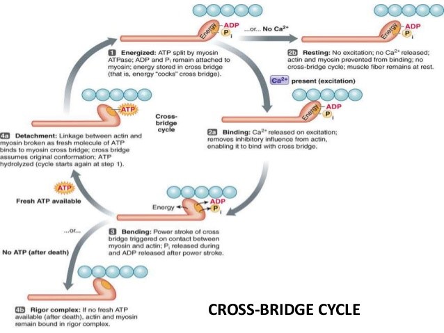
Myosin head pulls actin via ATP hydrolysis (ATPase) = contraction
SKELETAL
Pi release allows myosin head to move towards M line, pulling actin along with it.
Each movement pulls filaments approx. 10nm toward M line – this movement KA Powerstroke