Outline the physiology of the parasympathetic nervous system using the following headings:
the anatomical origins and target organ(s) (45% of marks). Responses of the organ(s) are NOT required.
nerve fiber classification and characteristics (20% of marks)
receptor types, locations and neurotransmitters (35% of marks
61% of candidates passed this question.
This question was best answered using the structure provided and ensuring the level of detail linked to the % of marks to be gained. It could be answered with the following simple information if presented with appropriate linking.
Craniosacral outflow.
CN III – eye,
CN VII – parotid gland
CN IX -submandibular glands,
CN X – cardiac, pulmonary and abdominal organs;
Sacral Outflow
S2-4 = pelvic and sex organs.
Preganglionic nerve fibres are long type B, partially myelinated and synapse on a ganglion close to the target organ. Post synaptic nerves are short type C unmyelinated.
Preganglionic NT is ACh onto N2 ACh receptor which is a ligand gated ion channel allowing for Na and K movement. Post ganglionic NT is ACh onto Muscarinic receptors which are G protein coupled.
M1, M4 and M5 are in the CNS (Gq, Gi, Gq respectively)
M2 – heart and lung (Gi – inhibition of AC and decreased cAMP)
M3 stomach, gut and other organs (Gq – IP3/PLC).
17A01: Exam Report
Outline the anatomy and physiology of the parasympathetic nervous system.
32% of candidates passed this question.
An efficient way to answer this question was to describe the anatomy and physiology of both cranial and sacral sections together. High scoring answers included an outline of the relevant nerves, the various ganglia, neurotransmitters and physiological effects. Some candidates described the cellular basis of Nicotinic, Muscarinic and M1-M5 receptors which didn’t attract marks.
14B04: Exam Report
Outline the anatomy and physiology of the parasympathetic nervous system.
0% of candidates passed this question.
Generally there was a lack of detailed knowledge, incorrect facts and at times confusion between the sympathetic and parasympathetic nervous system functions. A lack of anatomical detail was common (the origin of preganglionic cell bodies was not described clearly, and parasympathetic ganglia were not often named and located). It was expected an answer would mention the central role of Acetylcholine as a neurotransmitter at preganglionic and post ganglionic neurons in the parasympathetic system. Target organs were identified correctly but the exact action was not specified e.g. pupillary constriction vs. dilatation, GI sphincter/bladder – contraction vs. relaxation. Detail concerning receptor physiology was not required.
This is a question covering a core topic that no candidate passed. An overview of the arrangement and function of the autonomic nervous system is provided in several core physiology texts, including Ganong and Guyton.
M1i / 25B14 / 17A01 / 14B04: Anatomy & physiology of the parasympathetic nervous system
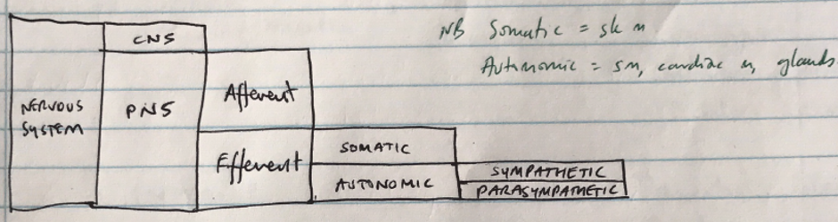
Parasympathetic NS = division of ANS responsible for restorative function
The ANS is the efferent pathway controlling the action of involuntary organs & tissues in order to maintain homeostasis
Parasympathetic Anatomy
Preganglionic Neurons
Myelinated B fibres
Long preganglionic fibres
Passes uninterrupted to ganglia near target organ
Craniosacral outflow
Preganglionic NT = ACh → acts on nicotinic receptors at ganglia
Postganglionic
Unmyelinated C fibres
Short due to location of ganglion near target organ
Postganglionic NT = ACh → acts on muscarinic receptors
Cranial Nerves
Preganglionic fibre origin
Ganglion
Postganglion Fibre Target
III
→ from oculomotor nucleus
→ ciliary ganglion
→ Eye
Ciliary m.
Iris sphincter
Pupil constriction
VII
→ Superior salivary nucleus
→ submaxillary ganglion
→ Submaxillary & sublingual
Salivary glands
↑saliva secretion
IX
→ Inferior salivary nucleus
→ otic ganglion
→ Parotid gland
↑saliva secretion
X
Vagus is the major component of parasympathetic outflow
→ Accounts for 75% of parasympathetic fibres
→ Dorsal nucleus of vagus (X) in medulla
→ Ganglia of visceral plexuses
→ Cardiac plexus, SA node, AV node, conducting system
↓chronotopy, chronotropy, conductivity
→ Pulmonary plexus
Bronchoconstriction
→ Gastric plexus
Stomach, liver, spleen
↑GI motility & secretions
Sphincter relaxation
↑peristalsis
Sacral nerves
S2, 3, 4 of spinal cord
→ Hypogastric plexus
→ Descending colon, rectum, bladder, uterus
Rectal contraction
Anus relaxation
Uterine relaxation
Contraction of detrusor m. in bladder wall
CHARACTERISTIC
PARASYMPATHETIC
Origin
Fibre length
Long preganglionic, short postganglionic
Preganglionic fibre
Myelinated B fibre
Ganglia location
Close to effector cells
Preganglionic NT
ACh
Ganglia receptors
Nicotinic
Postganglionic fibres
Unmyelinated C fibres
Postganglionic NT
ACh
Postganglionic receptor
Muscarinic
Function
Conserves & stores E.
ORGAN
ACTION
RECEPTOR
Heart
– inotropy
– chronotropy
– chronotropy
M2
Arteries
Dilatation
NB: not much effort on art/vein as for sympathetic NS