O1v / G4iii / 21A07 / 18A07: Outline the blood supply to the gastrointestinal system

21A07: Exam Report

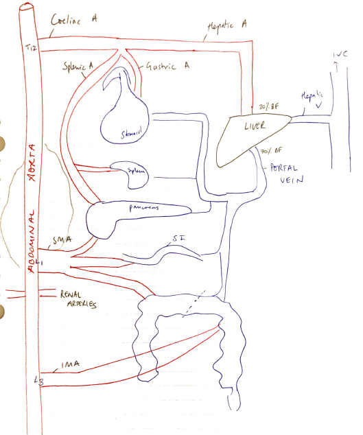
Outline the anatomy of the blood supply (arteries and veins) of the gastrointestinal system (oesophagus to anus)

48% of candidates passed this question.

This question was answered best if the main arteries and veins were discussed first and then their corresponding supply outline in reasonable detail. Very few candidates were able to achieve this. Listing the names of vessels with no context and in a random non-sequential order did not attract many marks. The physiology of the blood supply to the liver also did not attract marks.

18A07: Exam Report

Outline the blood supply to the gastrointestinal system (arteries and veins).

7% of candidates passed this question.

An outline of the blood supply from the oesophagus down to the anus was expected. Very few candidates knew the branches of the main 3 arteries and which portion of the gastrointestinal system they supplied. Concepts related to control of blood flow and autoregulation of blood flow were not asked and therefore marks were not awarded for this information.

O1v / G4iii / 21A07 / 18A07: Outline the blood supply to the gastrointestinal system

Splanchnic Circulation = blood flow to abdominal gastrointestinal organs → stomach, duodenum, SI, LI, liver, pancreas, spleen

Anatomy

  • 3 major vessels of ABDOMINAL AORTA (hiatus of diaphragm → Common Iliac A)

1. Coeliac Artery (T12)

  • Foregut (stomach → bile duct enters duodenum)
    • L) Gastric A
    • Splenic A
    • Common Hepatic A → L + R hepatic Arteries

2. SMA (L1)

  • Midgut (where bile duct enters duodenum → 2/3 across T colon)
    • Inferior pancreaticoduodenal A
    • Jejunal & ileal A
    • Middle colic A
    • Right colic A
    • Ileocolic A
  • SMA = largest splanchnic vessel
    • >10% CO
    • ∴emboli here are v. problematic

3. IMA (L3)

  • Hindgut (2/3 across T colon → rectum)
    • L) colic A
    • Sigmoid A
    • Superior Rectal A

Stomach

  • Very rich blood supply
    • Lesser curve
    • Greater curve
    • Fundus → Short gastric arteries (from splenic a.)
  • GASTRIC VEINS → PORTAL V.

Duodenum

  • Duodenal arteries (Coeliac Trunk + SMA)
  • Coeliac trunk → Sup. Pancreaticoduodenal A
  • SMA → inf. Pancreaticoduodenal A
  • DUODENAL VEINS → PORTAL V.

Jejunum & Ileum

  • SMA → SMV → PORTAL V.

Large Intestine

  • SMA → caecum, appendix, asc. colon, T colon
  • IMA → distal T colon, desc. colon, rectum
  • Superior + Inferior Mesenteric Veins → PORTAL V.

Spleen

  • Splenic a. → splenic V → joined by IMV → unites with SMV → PORTAL V.

Pancreas

  • Supplied by branches of tortuous SPLENIC A. which runs along sup. border of pancreas
  • Addit supply from + Inf. Pancreaticoduodenal A.
  • PANCREATIC V’s → SPLENIC V (joined by IMV) → unites SMV → PORTAL V.

Regulation of Circulation

  • At rest splanchnic BF = 30mL/100g tissue/min = 30% CO
  • Can vary 10mL (hypoperfusion) → 250mL (post-prandial)
  • ∴Highly adaptive BF regulation

Control of Splanchnic Circulation

  • INTRINSIC
    • Myogenic
    • Metabolic
  • EXTRINSIC
    • ANS
    • Humoral

Myogenic

  • Vessel response to Transmural P
  • ↑transmural P
  • Smooth m. contraction
  • VC
  • Maintains perfusion pressure

Metabolic

  • Metabolites which vasodilate accumulate ∴restoring/↑BF
  • ↑delivery of O2 washes these away → VC

ANS

  • All splanchnic circulation innervated by symp. fibres
  • Release NA → α adrenergic VC → redirect BF
  • Parasym. of vagus & pelvic nerves
  • Synapse post-ganglionic fibres in gut wall
  • ↑motility, ↑secretions, ↑BF
  • Activation M1 muscarinic → release NO → VD & ↑BF

Humoral

  • GI hormones released after meal ingestion
  • Gastrin & CCK = ↑intestinal BF