Describe the structure of the neuromuscular junction (NMJ), including the location of receptors and enzymes (a diagram may assist in your answer but is not required to achieve full marks) (30% of marks).
Explain the events that occur at the NMJ that lead to the onset and offset of a muscle/motor end plate action potential (a description of excitation/contraction coupling is NOT required) (70% of marks).
76% of candidates passed this question.
Diagrams were useful for many candidates, however others chose to provide a detailed description and also scored well. High scoring answers outlined the structure of the neuromuscular junction (NMJ) including all key functional elements.
This section required a clear and ordered explanation from the arrival of a presynaptic action potential via motor axon until the restoration of a resting membrane potential in the post-synaptic motor endplate. Many answers ceased their description following the binding of acetylcholine to the nicotinic receptor and degradation by acetylcholinesterase omitting the final steps in returning the motor end plate to its resting state. Common errors arose from confusion between skeletal and cardiac myocyte action potentials.
19A17: Exam Report
Explain the physiology of neuromuscular transmission.
60% of candidates passed this question.
Description of sequential events from axon conduction to detail at the neuromuscular junction was required. Well-constructed answers defined neuromuscular transmission, elucidated the structure of the neuromuscular junction (best done with a detailed diagram), described the central importance of acetylcholine, including synthesis, storage, receptors, and degradation. An ideal answer also described both pre-synaptic (e.g. voltage-gated calcium channels, exocytosis of vesicles) and post-synaptic events (acetylcholine receptors, end plate potentials, and the events that lead to excitation-contraction coupling in skeletal muscle).
L1ii / 25A14 / 19A17: Explain the physiology of neuromuscular transmission
Definition
NMJ = interface between nervous system & skeletal m.
NT = ACh
Skeletal m. innervated by Aα motor neuron
Aa = large diameter, myelinated, arise from anterior horn SC
At muscle, Aα axon divides into branches
Each axon forms a single junction with a muscle fibre
1 group muscle fibres innervated by 1 motor n. = MOTOR UNIT
Each Aα motor n. can innervate 1 – 2000 muscle fibres
But each muscle fibre only supplied by 1 Aα motor n.
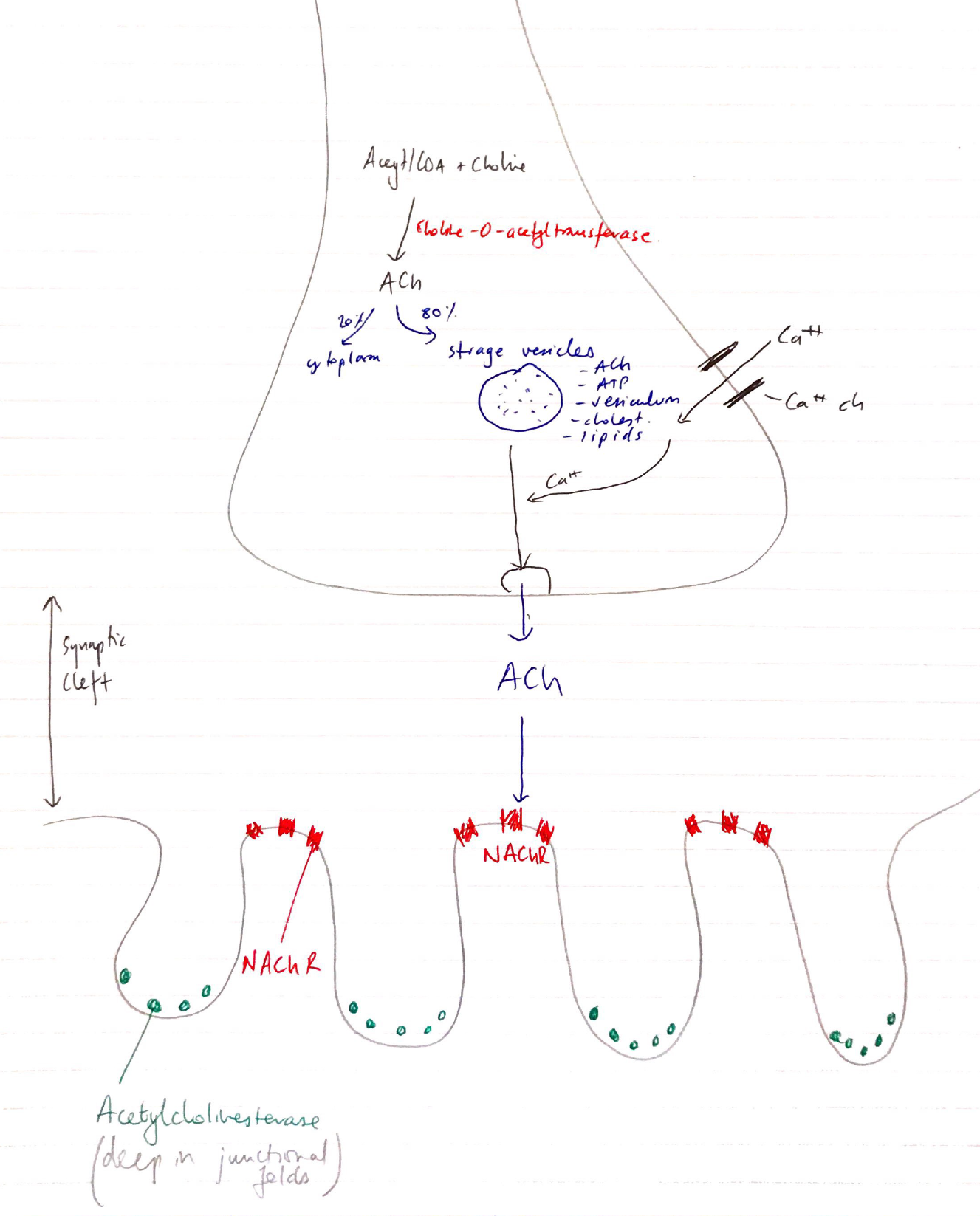
Anatomy
Aα motor n. approaches muscle → loses myelin & branches into ‘Terminal Buttons’
Muscle membrane opp Terminal Button is invaginated to form JUNCTIONAL FOLDS
NAChR is at the top of fold → Anti-cholinesterase enzymes located in valley of folds
Space b/w Motor End Plate & Terminal Button KAJUNCTIONAL CLEFT
ACh Synthesis & Release
Nerve cytoplasm: Acetyl CoA + choline → ACh
80% stored in vesicles, 20% stored in cytoplasm
ACh vesicles transported to NMJ
AP arrives at Terminal Button
Ca2+ influx at presynaptic terminal
Vesicle fuses with membrane
Releases contents into NMJ
Delayed opening K+ channel then restores membrane potential
NAChR & Events Leading To E-C-C
5 units: 2 α, β, γ
One ACh binds 2 x subunits (+ve cooperativity)
Channel opens = ↑ permeability to cations (Na, K, Mg)